

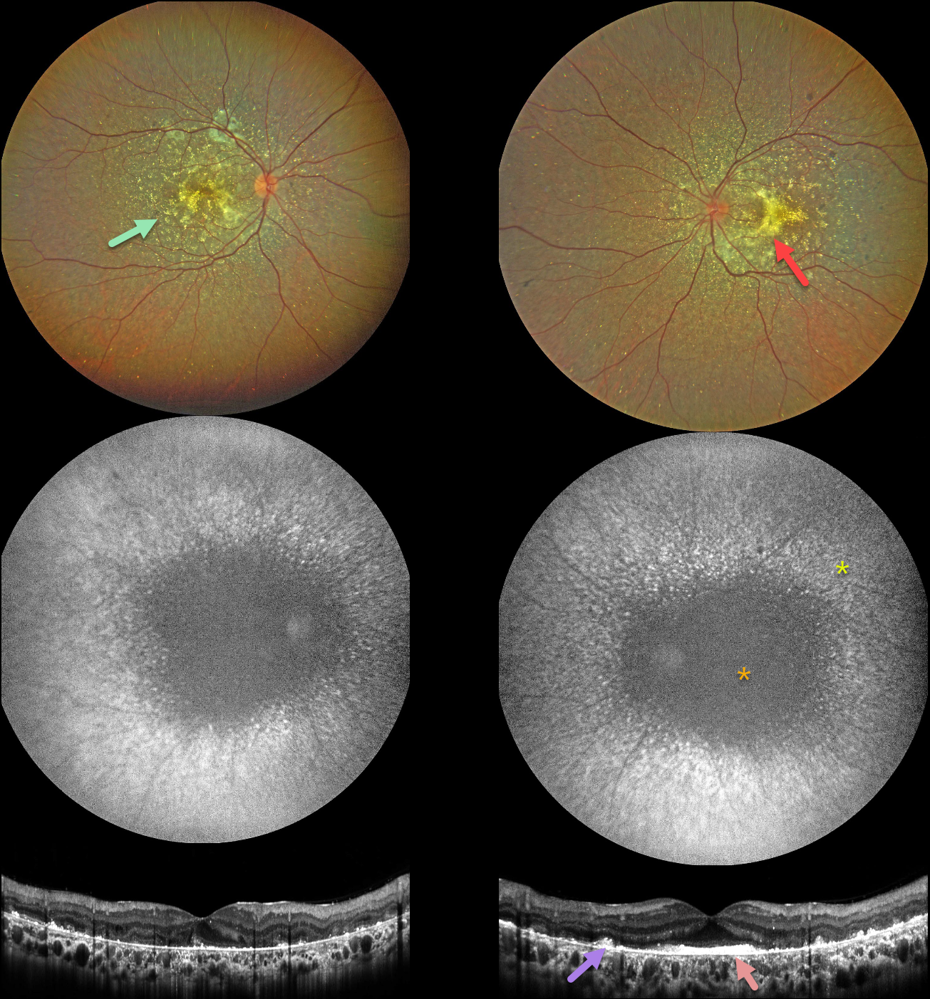
This 28YO Egyptian male reported blurry vision, nyctalopia, and photosensitivity. His great-grandmother was blind from an unknown etiology. Vision was 20/40 OD and 20/60 OS.
Anterior segments were notable for bilateral limbal crystalline deposits (not shown). Fundus imaging shows extensive subretinal crystalline deposits primarily in the posterior poles bilaterally, with areas of subretinal fibrosis.
Fundus autofluorescence (FAF) shows central hypo-FAF with radiating dots of more peripheral hyper-FAF.
OCT scanning shows hyperreflective dots overlying the RPE-Bruch’s membrane complex, with variable outer retinal and RPE loss. A placoid area of subfoveal hyperreflectivity is also noted OS.
Genetic testing revealed pathogenic heterozygous CYP4V2 mutations, consistent with Bietti crystalline dystrophy.
Learning Points:
Bietti crystalline dystrophy is an autosomal recessive ocular disorder that affects both the anterior and posterior segments. Clinical features include crystalline deposits in the retina, cornea, and rarely the crystalline lens, as well as retinal pigment epithelial clumping and atrophy.
Mutations in CYP4V2, which cause dysfunctional lipid metabolism, have been implicated in its pathogenesis. While there is no definitive treatment, anti-VEGF therapy can be administered for secondary macular neovascularization. See Saatci et al for a recent review (Clinical Ophthalmology 2023;17:953-967).



