

This 78YO hypertensive female presented with sudden vision loss in her left eye. Vision was 20/40 in her normal right eye and counting fingers in her left eye.
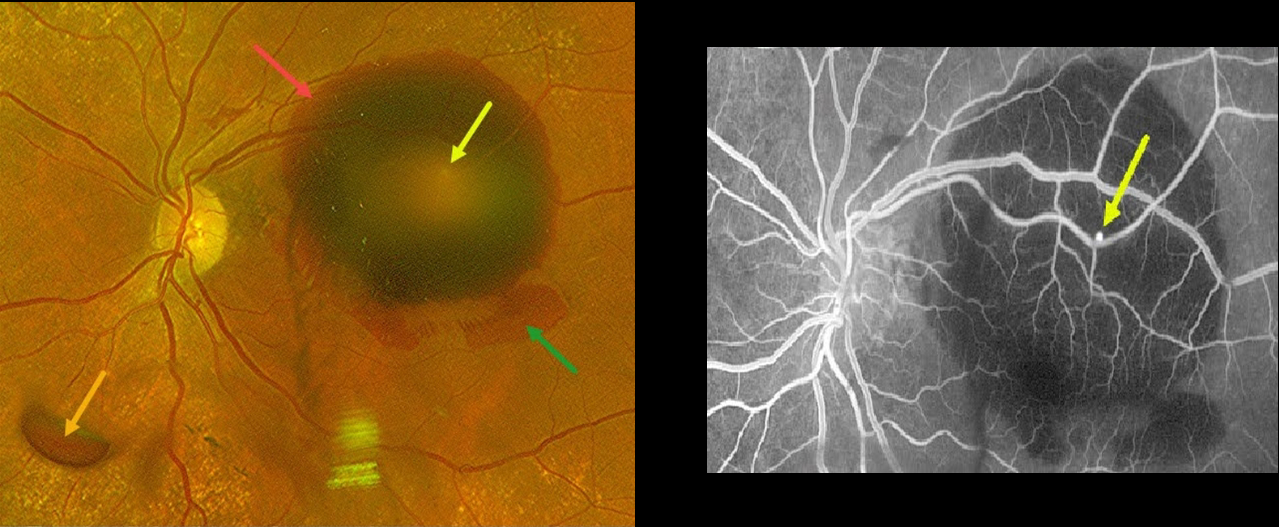
Optos color RG imaging shows multiple layers of blood, including a large submacular hemorrhage, intraretinal blood, and preretinal blood.
A tiny retinal arterial macroaneurysm is noted in the superotemporal macula clinically as well as on fluorescein angiography.
This case was submitted by Mohammad Abbas.
Learning Points:
A RAM is thought to occur in a weakened arterial wall from arteriosclerosis, and appears as a yellow-white dilated lesion along a retinal artery within the first 3 bifurcations from the optic nerve.
There are 2 distinct presentations, hemorrhagic and exudative. The hemorrhagic type often presents, as in this case, with pathognomonic multiple layers of blood. Acute macular hemorrhage can be treated with observation, anti-VEGF injections, or thermal laser.
Due to the thick submacular blood, vitrectomy with subretinal TPA was recommended. However, she declined surgery and is being followed for now.



