

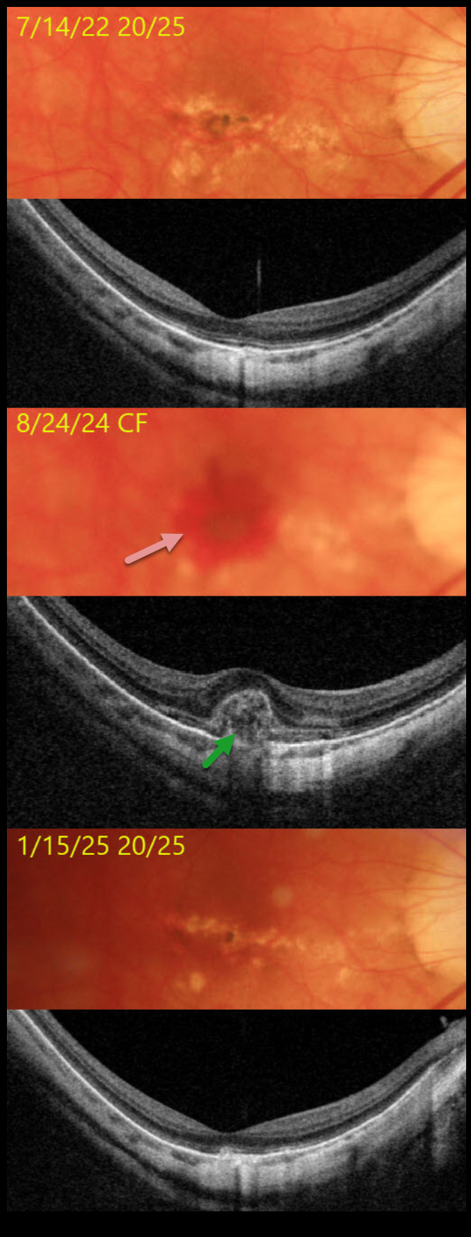
This 53YO highly myopic male was seen on 7/14/22 for a routine exam with 20/25 vision. Triton color imaging shows dry myopic degeneration, macular pigment loss, and clumping. Triton swept-source OCT shows variable loss of the outer retinal bands and diffuse choroidal thinning.
He returned on 8/24/24 with vision loss from a counting-fingers submacular hemorrhage. OCT shows hyperreflective subretinal blood elevating and extending into the outer retina. Following monthly intravitreal Avastin injections, vision improved to 20/25 on 1/15/25. All blood resolved, and the OCT mostly returned to its pre-blood appearance.
Learning Points:
In our experience, myopic macular neovascularization (MNV) presents quite differently from neovascular AMD. Significant subretinal blood (as in our patient), subretinal fluid, or intraretinal fluid are rarely seen. Often, an active MNV will present as a new dot of subretinal blood or pigment. Occasionally, there will just be symptomatic vision loss or metamorphopsia without clinical or OCT findings. Anti-VEGF therapy is effective, and these lesions can often be managed with PRN therapy, which is what we recommended for our patient (Sakata et al, Retina 2023;43:1863-1871).



