

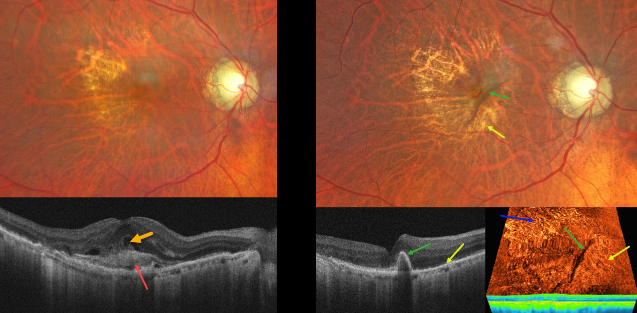
This 82YO male presented with recent vision loss in his right eye. Vision was 20/200.
Color photography shows a poor foveal reflex with superotemporal geographic atrophy. OCT scanning shows subretinal hyperreflective material (SHRM) with overlying retinal fluid. A possible bacillary layer detachment is also noted. About 6 weeks following a single intravitreal anti-VEGF injection, vision improved to 20/50. A new RPE tear is noted, with the bed of the tear inferonasal to the retracted hyperpigmented RPE. Despite persistent flow signals on OCT angiography, there is no fluid on structural OCT. The SHRM has completely resolved. A treat-and-extend anti-VEGF regimen was begun.
Learning Points:
A tear in the RPE is a fairly common finding in RPE detachment (PED) associated with type 1 macular neovascularization (MNV) located below the RPE. The neovascularization can spontaneously contract or involute following treatment. It then puts traction on the overlying RPE, which can then rip at the opposite edge of the PED. Like a cheap window blind, the torn RPE retracts and scrolls towards the MNV. This leaves a depigmented region where the RPE was and is now absent, along with a pigmented, relatively straight subretinal lesion from the scrolled RPE edge.
SHRM is an OCT finding that is invisible clinically. This material, located between the neurosensory retina and RPE, is thought to consist of fluid, fibrin, blood, and other fibrovascular tissues. SHRM is a biomarker of active disease that decreases with successful anti-VEGF therapy (Willoughby et al, Ophthalmology 2015;122:1846-1853). Persistent SHRM is associated with an increased incidence of scar formation and worse vision.
It is impossible to determine whether the anti-VEGF injection alone or in combination with the RPE tear caused such a rapid resolution of the SHRM. Fortunately, our patient’s vision remained quite good since the RPE tear just spared the foveal center.



