

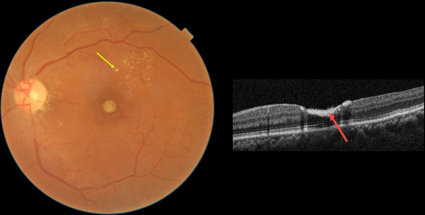
This 54YO female presented with 3 weeks of vision loss in her left eye. Vision was 20/20 in her normal OD and 20/200 OS.
Fundus photography shows devitalized yellow prefoveal blood, some lipid flecks centered in the superotemporal macula, and a tiny fibrosed superior retinal arterial macroaneurysm (RAM).
OCT scanning through the fovea confirms the sub-internal limiting membrane (ILM) location of the hyperreflective blood.
This case was submitted by Asma Samsudeen and Ashish Sharma.
Learning Points:
A RAM is thought to occur in a weakened arterial wall from arteriosclerosis, and appears as a yellow-white dilated lesion along a retinal artery within the first 3 bifurcations from the optic nerve.
There are 2 distinct presentations, hemorrhagic or exudative, and our patient has features of both. Although she reported only a several-week history of vision loss, the retinal findings suggest a much longer course.
The devitalized blood usually takes months to turn yellow, and was likely much larger in extent originally. The blood elsewhere has largely resolved. There was also likely macular thickening and possibly subretinal fluid, which spontaneously resolved, leaving behind residual peripheral macular lipid.
Observation was recommended, and we expect her vision to significantly improve, as the retina appears structurally fairly normal on OCT deep to the blood. Subretinal blood, especially devitalized, is toxic to the outer retina, which is why removal and/or displacement of thick subretinal blood is often recommended. Blood in the sub-ILM or preretinal space does not seem to damage the retina, so it can more safely be observed (see Bloom and Spaide, Retinal Cases 2022;16;401-402).



