

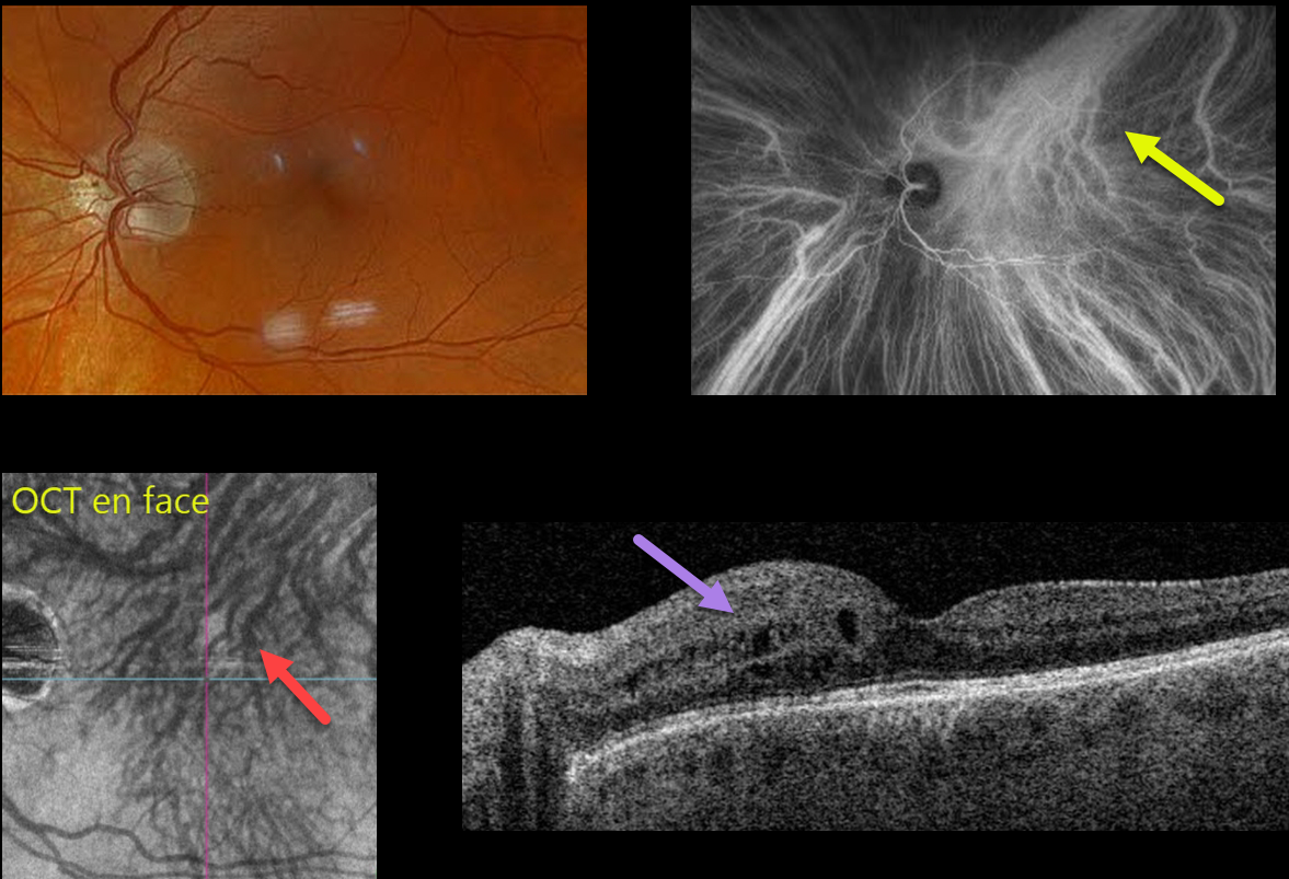
This 59YO male has been followed for the past year with asymptomatic peripapillary pachychoroid syndrome (PPS) in his left eye. Vision is stable at 20/25 OU.
Optos color RGB imaging shows a normal left macula. Indocyanine green (ICG) angiography is normal OD (not shown), but reveals a dilated vortex system superotemporally that drains nearly the entire macula, along with a strikingly similar appearance from the OCT choroidal en face.
Triton swept-source OCT was normal OD (not shown), but shows mild nasal thickening with cysts in the inner nuclear layer OS.
Learning Points:
Originally described by Phasukkijwatana et al in 2018 (Retina 2018;38:1652-1667), PPS presents with nasal macular intraretinal and subretinal fluid. These eyes can look very similar to those of patients with optic pit maculopathy, but without the pit.
Additional findings include a thicker nasal than temporal macular choroid, choroidal folds (75% of cases), shorter axial lengths (<23mm), and hyperopia. There is no angiographic leakage despite the OCT fluid. This fluid will often spontaneously wax and wane. Symptomatic fluid can successfully be treated with photodynamic therapy.
Intervortex venous anastomoses are common in pachychoroid-related disorders (Spaide et al Retina 2021;41:997-1004). In this case, the choroidal anastomosis and dilated superior vortex system were beautifully imaged on both ICG and en face OCT.
In our practice, we continue to find that en face imaging often provides a readily accessible, unique, and compelling perspective for a variety of pathologies, and we hope other clinicians will add this to their daily clinic routine.



