

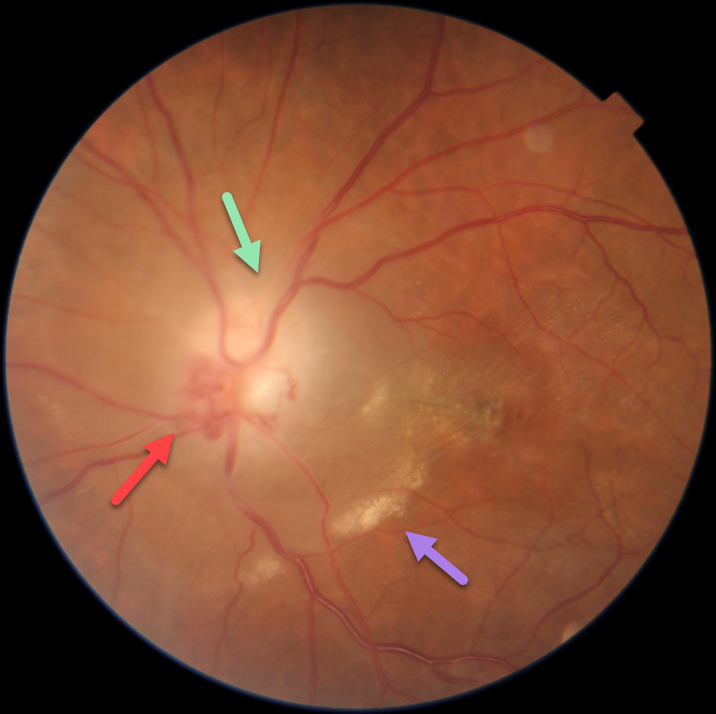
This 40YO female presented with vision loss in her left eye for 7 months. Vision was 20/20 in her healthy right eye and hand motion in her left eye.
Fundus photography shows an ill-defined, somewhat pale left nerve with peripapillary fluid, subretinal lipid, and foveal pigmentary changes. Disc collaterals are noted. Externally, the left globe was proptotic and displaced down and out (not shown).
MRI scanning was highly suggestive of an optic nerve glioma, and she was immediately referred for neurosurgical evaluation.
This case was submitted by Asma Samsudeen and Ashish Sharma.
Learning Points:
An optic nerve glioma is a benign, slow-growing tumor most commonly seen in children and associated with neurofibromatosis type 1. This is in contrast to a glioblastoma, an aggressive malignant tumor that is more common in adults. Unfortunately, we do not have a definitive diagnosis since our patient was subsequently lost to follow-up.
Collateral vessels are dilated, pre-existing vessels that bypass an occlusion. These are most commonly seen at the posterior pole following retinal venous occlusions, but can also occur at the optic disc with optic nerve meningiomas or gliomas. The endothelial tight junctions are intact, so they don’t leak on angiography. This helps differentiate them from neovascularization, which does leak.



