

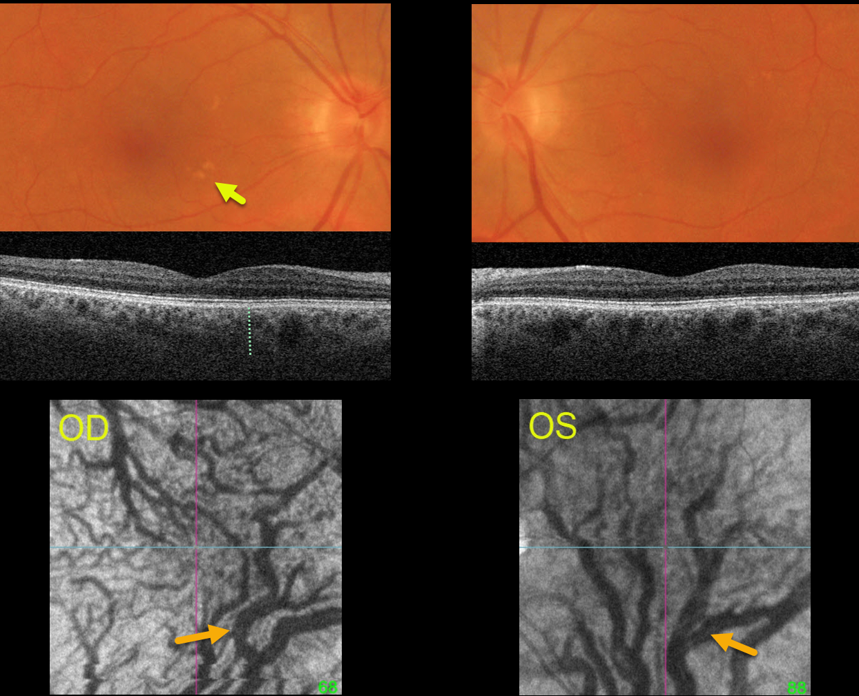
This healthy 67YO female was examined for a symptomatic posterior vitreous detachment in her right eye. She was previously visually asymptomatic, and vision was 20/25 OU.
Optos color RGB imaging shows several large irregular drusen in the nasal right macula and faint drusen in the superotemporal left macula.
Triton swept source OCT shows a relatively thickened choroid for the patient’s age, along with large choroidal vessels. En face imaging through each choroid shows marked vortex dilation bilaterally. Each macula, particularly in her left eye, is drained by the inferior vortex system.
Learning Points:
Pachychoroid, meaning “thick” choroid, is secondary to large dilated Haller vessels. Increased oncotic pressure from these dilated vessels can cause RPE dysfunction, serous detachment, and macular neovascularization.
Another, more subtle finding is pachydrusen. Unlike typical age-related drusen that are pinpoint or round and centered in each macula, pachydrusen are fewer in number, more ectopic, and often have an irregular contour (Spaide, Retina 2018;38:708-716).
The choroid is typically thinned with AMD, but eyes with pachydrusen have thicker choroids. Although our patient’s macular clinical findings were quite subtle and the OCT B-scans were suggestive of pachychoroid, her en face images most dramatically show the choroidal pathology.



