

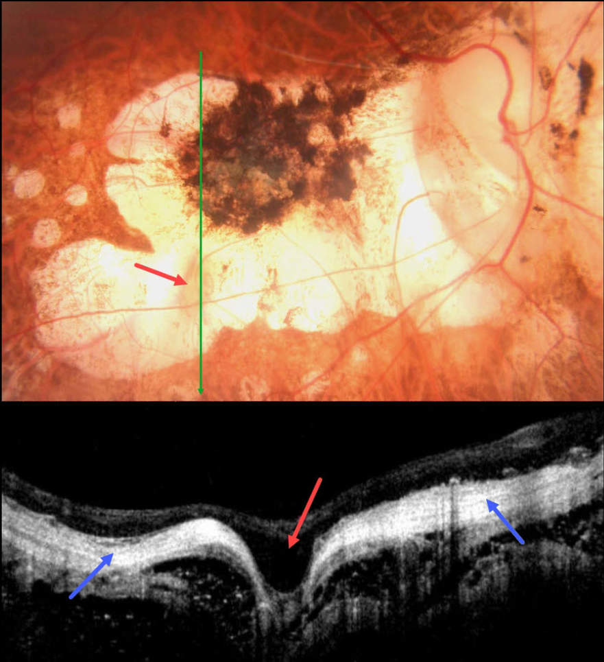
This 58YO male with a history of degenerative myopia presented with 6 months of vision loss in his right eye. He was blind in his left eye following vitrectomy surgery 7 years earlier. Vision was counting fingers OD.
Color photography shows severe variably pigmented atrophic myopic degeneration. An area of orange coloration is seen within the larger area of atrophy inferiorly.
On OCT, this area appears as a focal staphyloma. The overlying retina is draped across the staphyloma. The choroid is so thin as to be invisible on OCT, with a hyperreflective underlying sclera. The orbital tissue is well seen posterior to the sclera.
Learning Points:
Degenerative myopia is a major cause of worldwide visual impairment. Due to the stretching of the posterior retinal structures, there is an increased risk for numerous findings, including atrophic retinal and choroidal thinning, posterior staphyloma, macular schisis/traction, and macular neovascularization (see Ohno-Matsui et al for a recent review, Invest Ophthalmol Vis Sci 2021;62(5);5)..
Initially described by Freund et al (Arch Ophthalmol 2003;121:197-204), intrachoroidal cavitations are most commonly found in high myopes as a peripapillary yellow-orange subretinal lesion. It is difficult to determine whether our patient has a cavitation above the staphyloma.



