

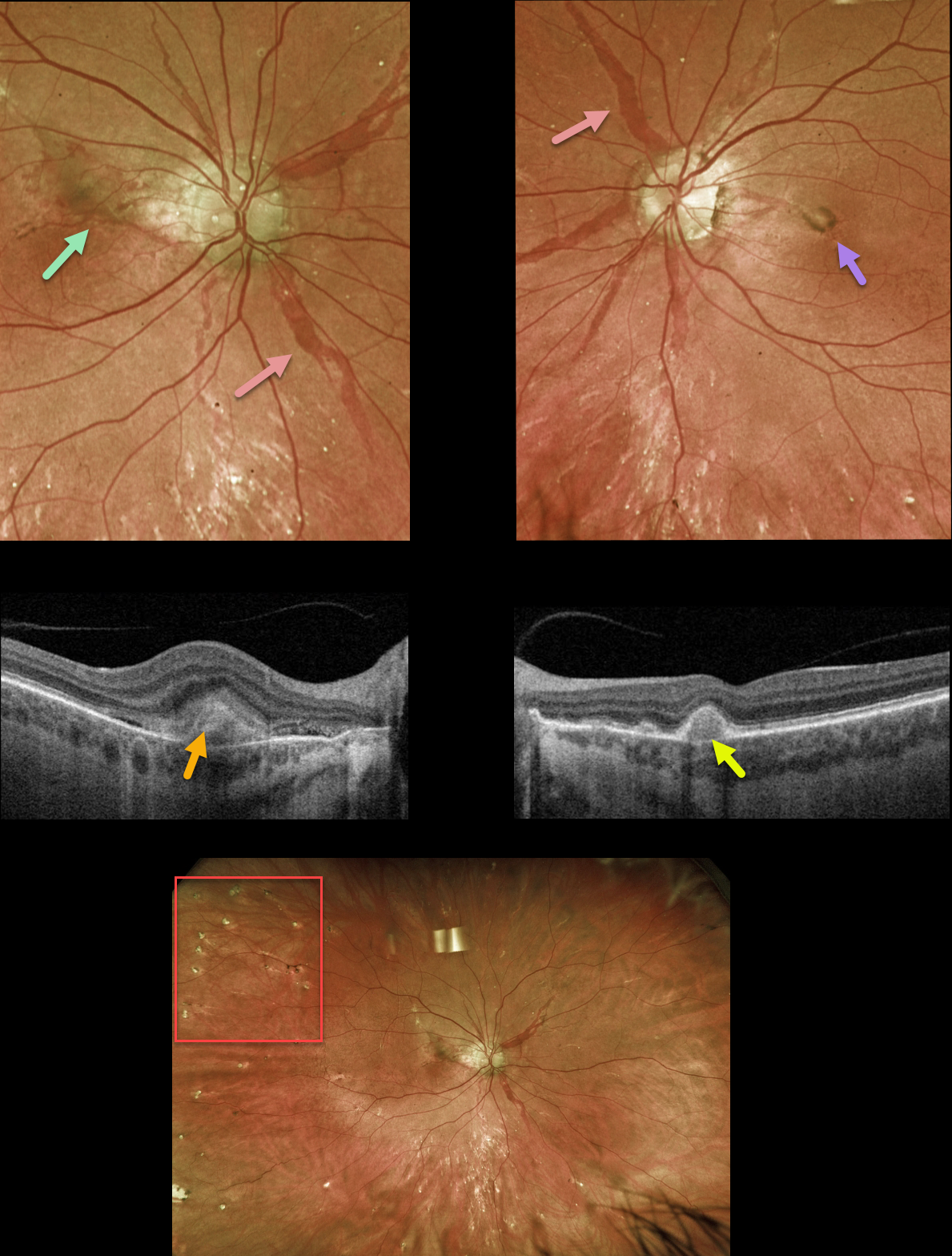
This 42YO female with a known history of pseudoxanthoma elasticum (PXE) presented with new central flashes in her right eye. Vision was 20/25 OD and 20/30 OS.
Optos color RG imaging shows large, irregularly dilated’ angioid streaks’ radiating outwards from each optic nerve. A pigmented macular neovascularization (MNV) with some subretinal blood is noted in the superior right macula, and a small inactive MNV is noted in the left nasal fovea. Innumerable small subretinal comet-shaped lesions (‘comet rain’) are noted.
Swept-source OCT through the right MNV shows a hyperreflective, thickened type 2 lesion (above the RPE). OCT through the left MNV shows a small, nodular, hyperreflective type 1 lesion (below the RPE).
Learning Points:
Angioid streaks are orange-yellow, irregular, crack-like dehiscences in Bruch’s membrane associated with atrophic degeneration of the overlying RPE. They are often missed clinically because their color and orientation blend with the overlying retinal vessels.
Our patient’s lesions are about the most dramatic and red angioid streaks we’ve ever seen and are pathognomonic for PXE, but less extensive streaks can also be seen in sickle cell and age-related macular degeneration.
The comet lesions are also pathognomonic for PXE. These lesions develop from the outer retina as a neurodegenerative process and strongly resemble the hyporeflective core with a surrounding hyperreflective rim found in outer retinal tubulation (see Murro et al., Graefe’s 2018;256:1605-1614).
These patients are at significant risk for macular neovascularization (MNV), as occurred in each of our patients’ eyes. Intravitreal Avastin therapy was started in her right eye. The left MNV will be observed, as it involuted spontaneously without prior symptoms.



