

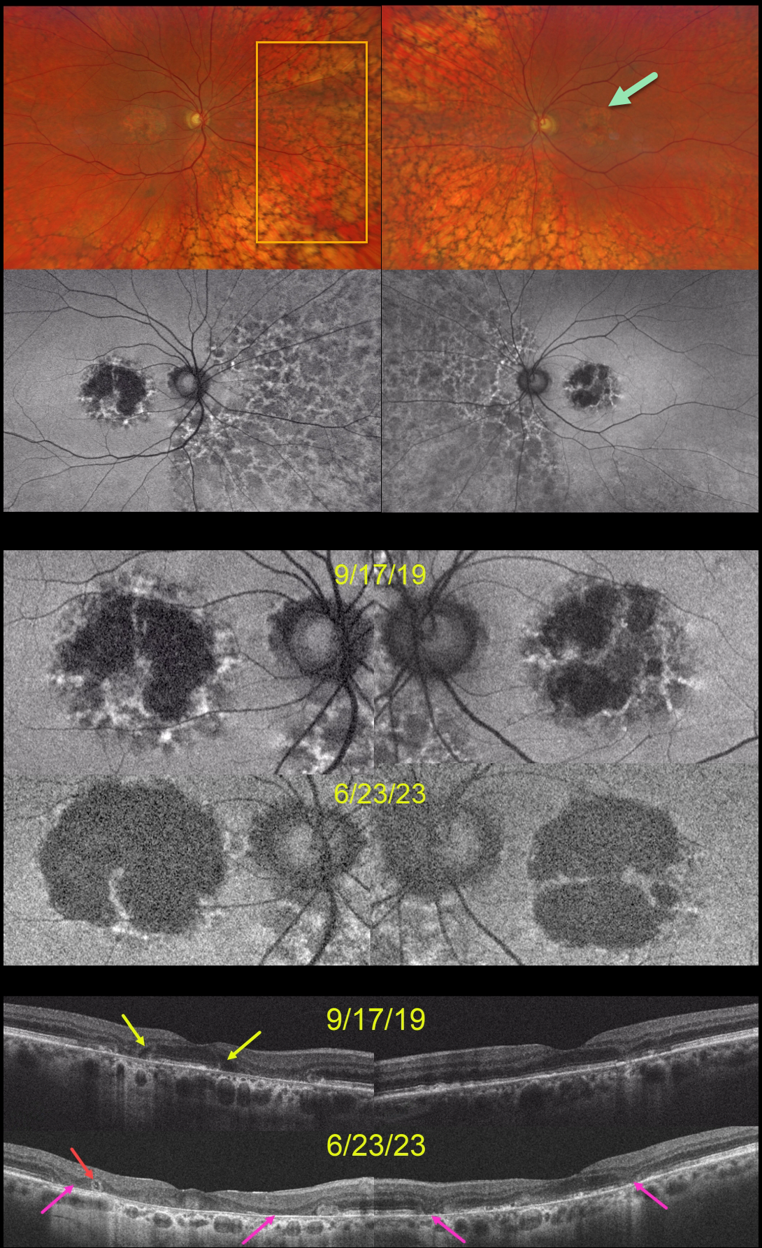
This 59YO was examined on 9/17/19. She was taking Elmiron (pentosan polysulfate sodium, PPS) for 17 years for interstitial cystitis. Vision was 20/40 OD and 20/30 OS.
Optos color RG imaging shows bilateral central-sparing macular atrophy (MA) and extensive, posterior peripheral reticular degeneration of the RPE (PRDRPE).
Fundus autofluorescence shows hypo-FAF from the MA with surrounding punctate and linear areas of hyper-FAF. Based on these findings, she decided to stop taking Elmiron.
When last examined on 6/23/23, vision dropped to 20/800 OD and was relatively stable at 20/40 OS. The MA progressed bilaterally clinically and on FAF and OCT. Hyporeflective OPL degeneration (wedge defects) is noted on the initial 9/17/19 OCT. On the 6/23/23 OCTs, an area of outer retinal tubulation (ORT) is seen temporally OD, and progressive outer retinal and RPE atrophy are noted bilaterally.
Learning Points:
Elmiron was approved by the FDA in 1996 for the treatment of interstitial cystitis. Recently, a unique PPS retinopathy has been described. Toxicity seems to develop over many years and can mimic more common disorders, including age-related macular degeneration and macular dystrophies.
Peripapillary hypoautofluorescence, more densely packed macular autofluorescent changes, and earlier central macular involvement suggest PPS toxicity over other causes (see Barnes et al Ophthalmology Retina 2020;4:1196-1201), including maternally inherited diabetes and deafness (MIDD).
ORT is often noted overlying inactive macular neovascularization with ongoing anti-VEGF therapy and should not be confused with exudative fluid or cysts, which lack a hyperreflective border. The outer hyperreflective band likely represents inner segment mitochondria undergoing fission and translocation towards the nucleus (Litts et al, Retina 2018;38:445-461).
ORT, initially described by Zweifel et al (Arch Ophthalmol 2009;127:1596-1602), is a neurodegenerative condition of the photoreceptors and Muller cells associated with outer retinal and retinal pigment epithelium atrophy, including advanced AMD and inherited retinal diseases.
The hyper-FAF surrounding the initial MA is more typical for PPS toxicity, and progressive atrophy is not uncommon even with stopping therapy (Jung et al, JAMA Ophthalmol 2023;141:260-266). However, in our experience, PPS usually produces more widespread macular findings. Given the PRDRPE, the MA could also be age-related. Or maybe our patient had baseline age-related MA superimposed on Elmiron toxicity?



