

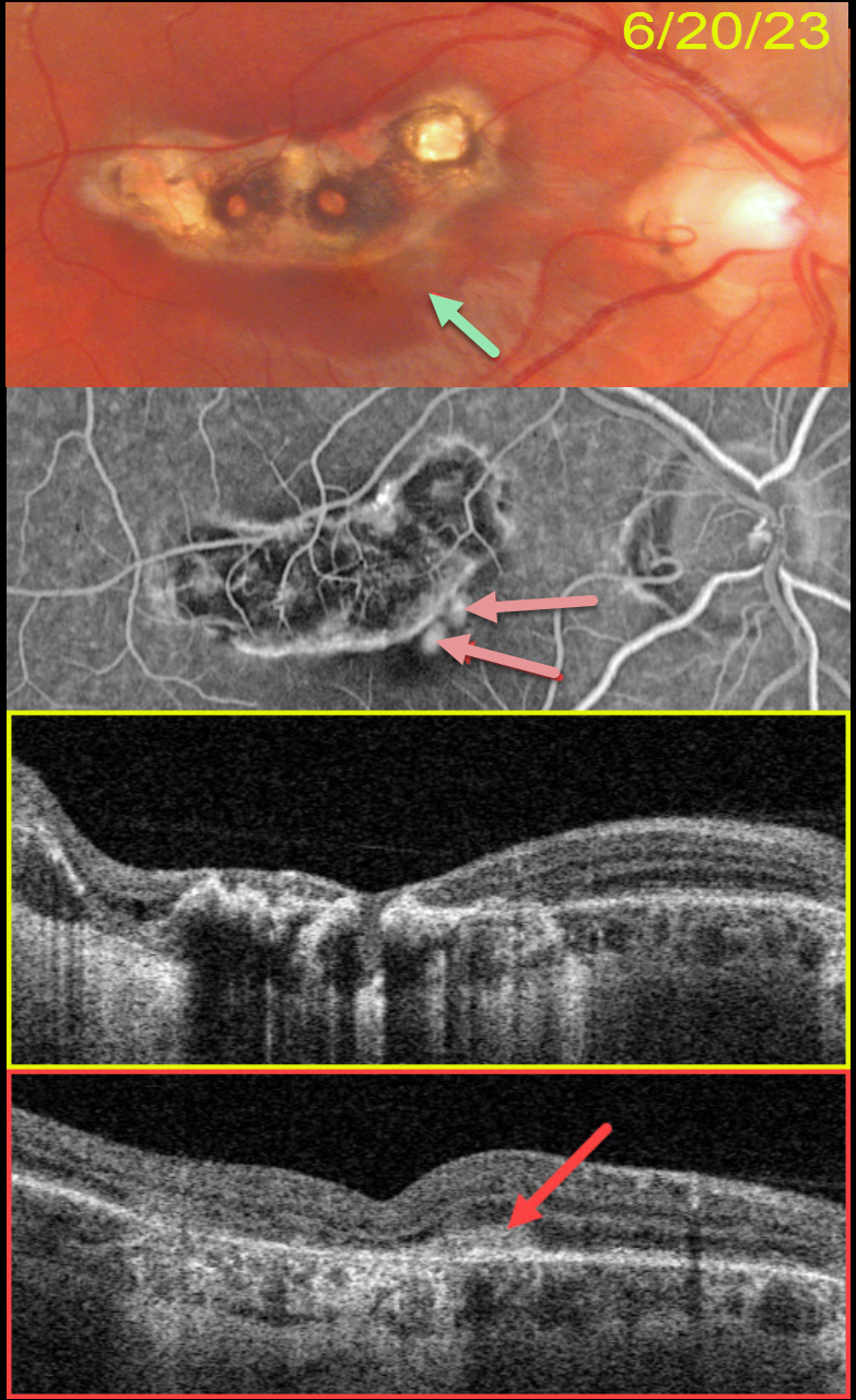
This 22YO male presented with several days of central blurred vision in his left eye. There was no prior ocular or medical history. Vision was 20/25 in his normal right eye and 20/25 in his left eye.
Color photography shows a variably pigmented comma-shaped colobomatous scar in the superior macula. A tiny depigmented scar is noted along its inferonasal edge. OCT scanning through this lesion showed no fluid (not shown).
He returned about 2 months later, complaining of increasing vision loss in his eye. Vision had decreased to 20/400. A faint area of subretinal pigment is now noted.
Optos fluorescein angiography shows leaking macular neovascularization (MNV) within this region. Swept-source OCT confirms a type 2 MNV (located above the RPE).
Intravitreal Avastin was injected.
Learning Points:
Virtually any disorder that affects the RPE-Bruch membrane layer can cause MNV, the most common being age-related macular degeneration, ocular histoplasmosis, idiopathic high myopia (lacquer cracks), trauma (choroidal rupture), and angioid streaks.



