

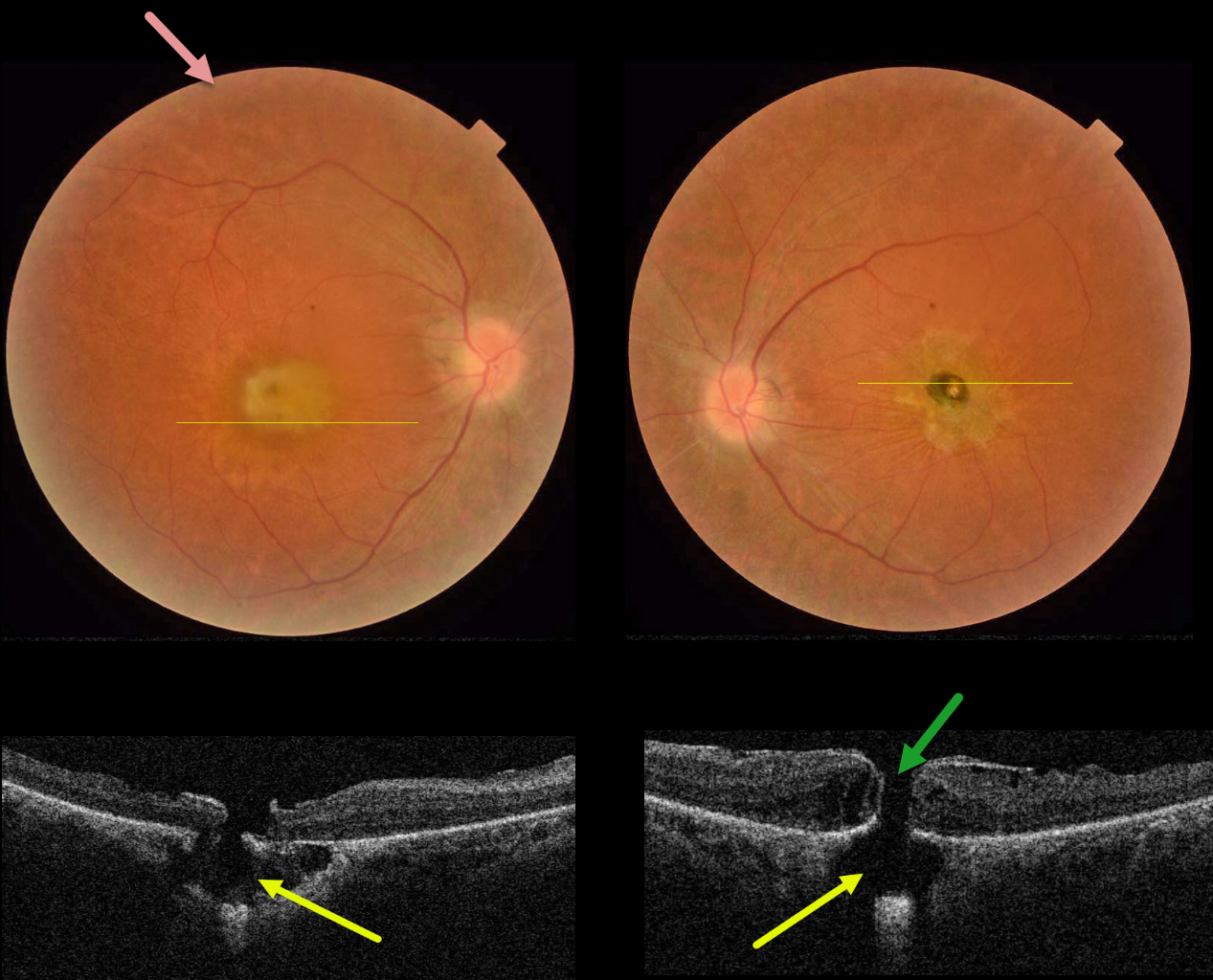
This 32YO female presented with a known history of severe vision loss from retinitis pigmentosa (RP). Her parents are first-degree cousins, and there was no family history of RP. Vision was hand motion OD and counting fingers OS.
Fundus photography shows bilateral irregular yellow central macular pigmentary changes, with a central hyperpigmented spot OS.
OCT scanning through each macula shows a full-thickness macular hole with an underlying defect in the RPE. Each macular hole is suspended over a choroidal cavitation.
Surgical repair was offered with appropriate counseling regarding the poor visual prognosis, and the patient elected to be observed.
Learning Points:
Initially described by Freund et al (Arch Ophthalmol 2003;121:197-204), intrachoroidal cavitations are most commonly found in high myopes as a peripapillary yellow-orange subretinal lesion.
Ornek and Ornek reported a patient with pathologic myopia and a unilateral macular hole communicating with a choroidal cavitation (Retinal Cases 2020;14:328-330).
Although macular holes can rarely be associated with RP (Jin et al, Retina 2008;28:610-614), to the best of our knowledge, ours is the first occurrence of RP with bilateral macular holes and underlying choroidal cavitations.



