

This 74YO male was seen for a routine eye examination. He told us he had a laser procedure on his right eye 18 years ago. Based on this image, what type of laser was it?
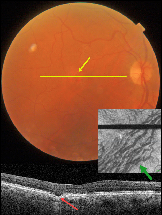
Fundus photography shows a normal macula except for an area of depigmentation in the temporal fovea.
Swept-source OCT through this area shows a small focal choroidal excavation. Although the choroid appears to have normal thickness, large outer choroidal vessels spanning virtually the entire choroid are noted nasally.
Choroidal en face OCT is markedly abnormal with dilated vessels and vortex anastomoses.
Our patient shows several pachychoroid-related findings, including a thick choroid for their age, large choroidal vessels, FCE, and vortex anastomoses. Our patient currently has asymptomatic pachychoroid despite having a ‘normal’ choroidal thickness on OCT.
So what type of laser did our patient have? Photodynamic therapy for central serous retinopathy, which has remained in remission since treatment in 2005
Learning Points:
Pachychoroid, meaning “thick” choroid, is secondary to large dilated Haller vessels. Increased oncotic pressure from these dilated vessels can cause RPE dysfunction, serous detachment, and macular neovascularization.
The choroidal thickness depends on numerous factors, including refractive error and age. A high myope’s choroid should be quite thin, but if it appears as a ‘normal’ thickness, it is thick for that eye. We also often see pachychoroid disease in elderly patients whose choroid is abnormally thickened for their age, but appears ‘normally’ thick when not accounting for this.
FCE, originally described by Jampol et al in 2006, is likely part of the pachychoroid spectrum, as it is usually associated with a thickened choroid and pachyvessels. The FCE can be observed, although symptomatic manifestations, including active central serous retinopathy or macular neovascularization, often require treatment. An excellent review is by Verma et al, Br J Ophthalmol 2021;105:1043-1048.
The choroidal venous system, as with the neurosensory retinal vasculature, usually respects the horizontal meridian. With pachychoroid disorders, these anastomoses develop, and in this case, the inferior vortex system drains nearly the entire macula.
For a wonderful review highlighting the concept of venous overload choroidopathy, see Spaide et al, Progress Retinal Eye Research 2022;86:100973.



