

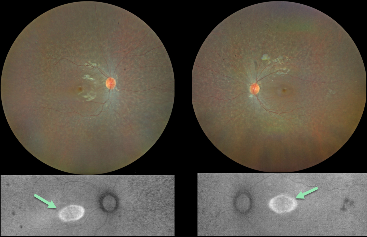
This 13YO girl has a history of congenital deafness, autism, and ADHD. Vision was 20/30 OU.
Color imaging shows bilateral and symmetrical moth-eaten pigmentary changes with an oval area of foveal hyper-autofluorescence.
Fundus autofluorescence (FAF) visualizes endogenous fluorophores, compounds that spontaneously fluoresce without an external dye.
The main fundus fluorophore is lipofuscin, which resides within the RPE lysosomes. The photoreceptor outer segments absorb some of the autofluorescent excitatory light, and thus normally diminish the FAF signal.
In inherited retinal diseases (IRDs), loss of outer segments unmasks this signal, leading to increased FAF. Many patients with an IRD will show a hyper-FAF ring between clinically normal and abnormal retina, which likely represents retina ‘at risk’, showing the junction of a functional and dysfunctional retina (Oishi et al, Int J Retina Vitreous 2019;5(S1);23).



