

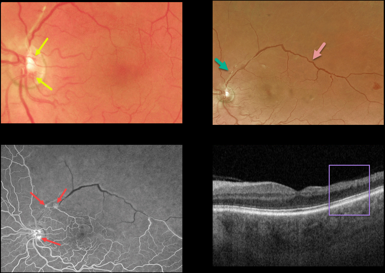
This 55YO male presented with relatively asymptomatic inferior field loss in his left eye.
Color imaging of the left nerve and macula shows a few small areas of disc neovascularization (NVD) and a fibrosed arteriole superiorly. Optos color RG imaging shows an irregularly dilated superotemporal major retinal vein and a featureless superior retina.
Fluorescein angiography shows severe retinal ischemia throughout the entire distribution of the branch retinal vein occlusion (BRVO) with leaking NVD and neovascularization elsewhere.
OCT scanning shows temporal inner retinal atrophy with the inner retinal layers replaced by a single hyperreflective band.
Scatter laser photocoagulation was recommended.
Learning Points:
In 1986, the Branch Vein Occlusion Study Group reported that scatter laser decreased the risk of vitreous hemorrhage from retinal neovascularization in BRVO, and suggested that laser should be applied once neovascularization develops (Arch Ophthalmol 1986;104:34-41).
Decades later, scatter laser remains the gold standard, although some may prefer to first stabilize neovascularization with anti-VEGF therapy prior to laser.



