

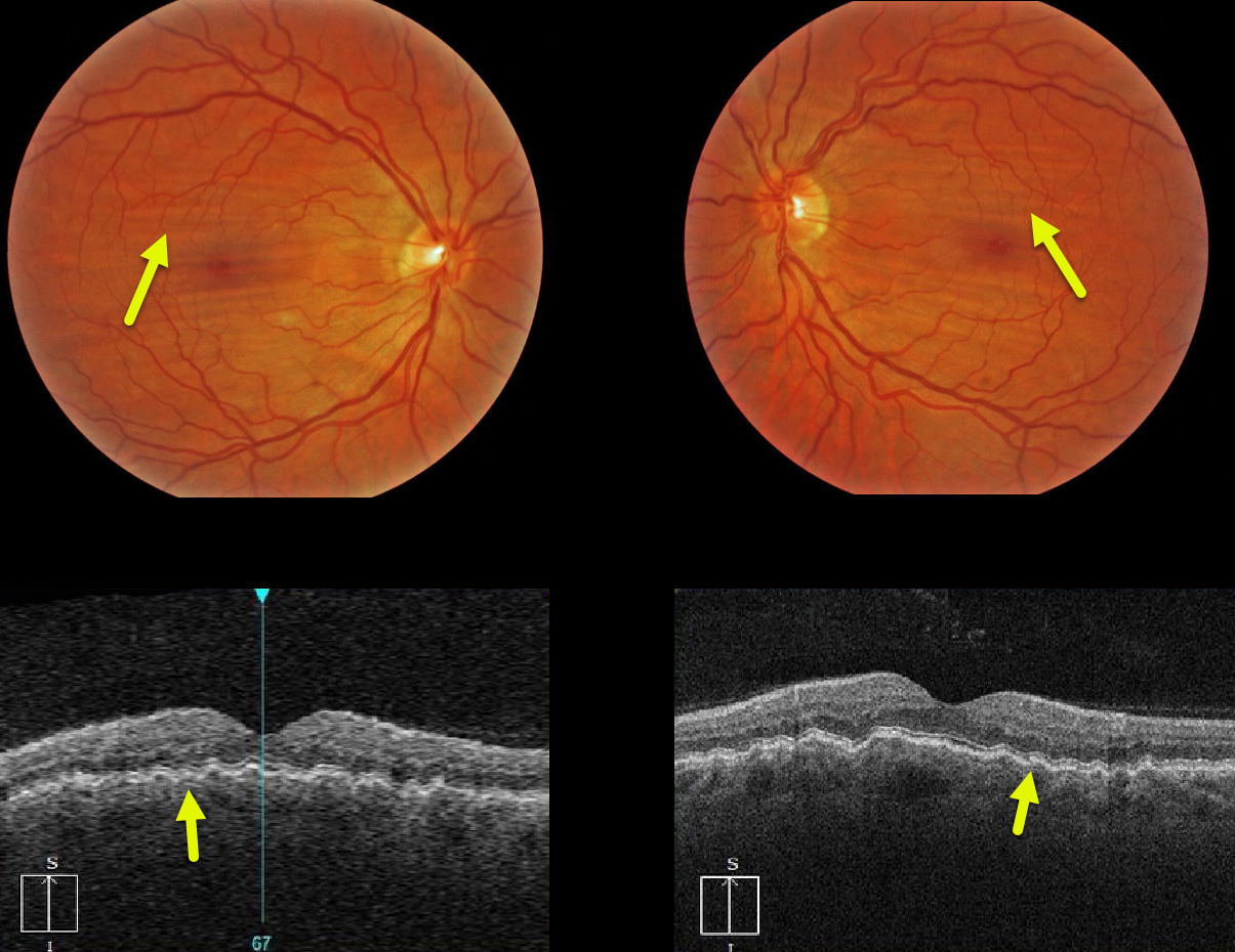
Our patient with mild hyperopia exhibits bilateral and symmetric idiopathic chorioretinal folds, shown on both the fundus photos and OCT images.
Learning Points:
Chorioretinal folds appear clinically as often subtle horizontal and oblique subretinal folds.
There are extensive intra- and extra-ocular causes for chorioretinal folds. Typical idiopathic chorioretinal folds require no workup and usually cause no symptoms.
When unilateral, an MRI is often indicated to rule out an orbital mass such as optic nerve meningioma.



