

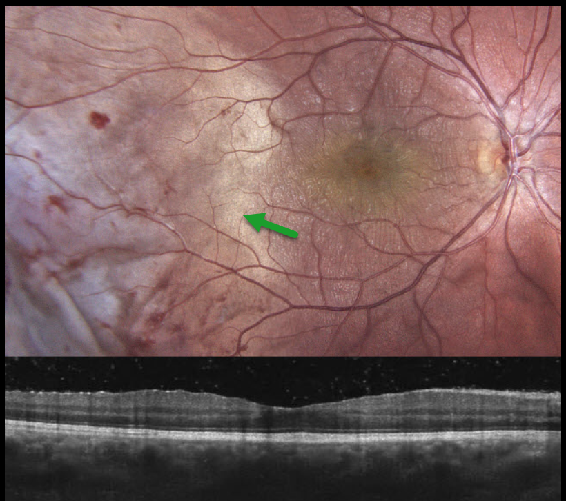
This 16YO male was chopping wood one day earlier when a branch recoiled and struck his right eye. Vision was 20/30 OD and 20/20 in his normal OS. The right anterior segment showed localized conjunctival chemosis with a subluxed crystalline lens.
Color photography shows temporal commotio retina extending into the temporal macula, along with scattered retinal hemorrhages. Although there appear to be fine inner macular striae, OCT scanning is fairly normal. The patient was referred for further management.
Learning Points:
Commotio retinae is caused by direct, blunt eye trauma. The outer retinal whitening is due to shearing of the photoreceptor outer segments. The term “Berlin’s edema” is a misnomer and should not be used, as the retina is not edematous. The acute white retinal changes usually resolve within several weeks. With resolution, the fundus appearance can normalize. With more severe injury, outer retinal loss can allow RPE cells to migrate into the retina, giving a pseudo-retinitis pigmentosa appearance. Patients can also develop retinal breaks and detachment.



