

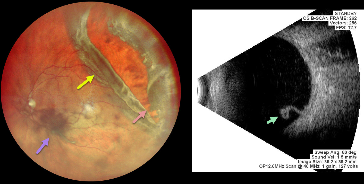
This 56 YO male presented with 4 days of vision loss in his left eye. He gave a history of multiple prior retinal reattachment surgeries in his right eye. Vision was 20/400 in his OD and 20/20 in OS.
Pseudocolor SLO imaging shows a superotemporal rhegmatogenous retinal detachment (RRD) from a giant retinal tear (extends over 3 clock hours) with mild vitreous hemorrhage. The anterior portion of the tear has multiple jagged edges, and the posterior flap has folded over onto itself. The macula is attached clinically and on B-scan ultrasonography, which shows the mobile posterior flap folded over onto itself. Emergency vitrectomy was recommended, but unfortunately, he was immediately lost to follow-up.
Learning Points:
A vitreous hemorrhage will often prevent visualization of an acute RRD. Ultrasonography is often utilized to confirm the presence of a detachment. In our patient, the B-scan also beautifully imaged the mobile posterior flap of the giant tear. This pathognomonic appearance would alter the surgical planning in the case of opaque media.



