

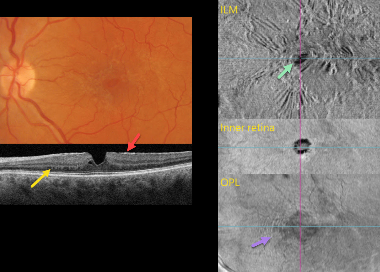
This 56YO female was referred for asymptomatic retinal findings. Vision was 20/20.
Multimodal imaging shows numerous pucker-related findings.
Triton color photography shows a diffuse macular pucker with a central teardrop-shaped pseudohole.
Triton swept-source OCT B-scanning shows an adherent hyperreflective epiretinal membrane, a vertical foveal contour, and trace adjacent outer plexiform splitting. The nasal outer plexiform layer (OPL) contains numerous peg-like hyperreflective lesions that extend into the outer nuclear layer.
En face OCT of the retinal surface shows variable striae and distortion from the pucker, along with the hyporeflective pseudohole. En face OCT through the inner retina shows the irregular pseudohole. En face of the OPL, through the peg-like lesions noted on the B-scan, shows a hyperreflective fingerprint.
Learning Points:
The ‘fingerprint sign’ is a unique OCT en face finding, described by Griffin et al (Retina 2021;41:381-386), appearing as concentric, fingerprint-like waves of Henle’s layer underlying epiretinal membranes. The authors felt that the undulating OPL creates hyperreflective lines when oriented perpendicular to the incident OCT beam and hyporeflective lines when oriented parallel to it.



