

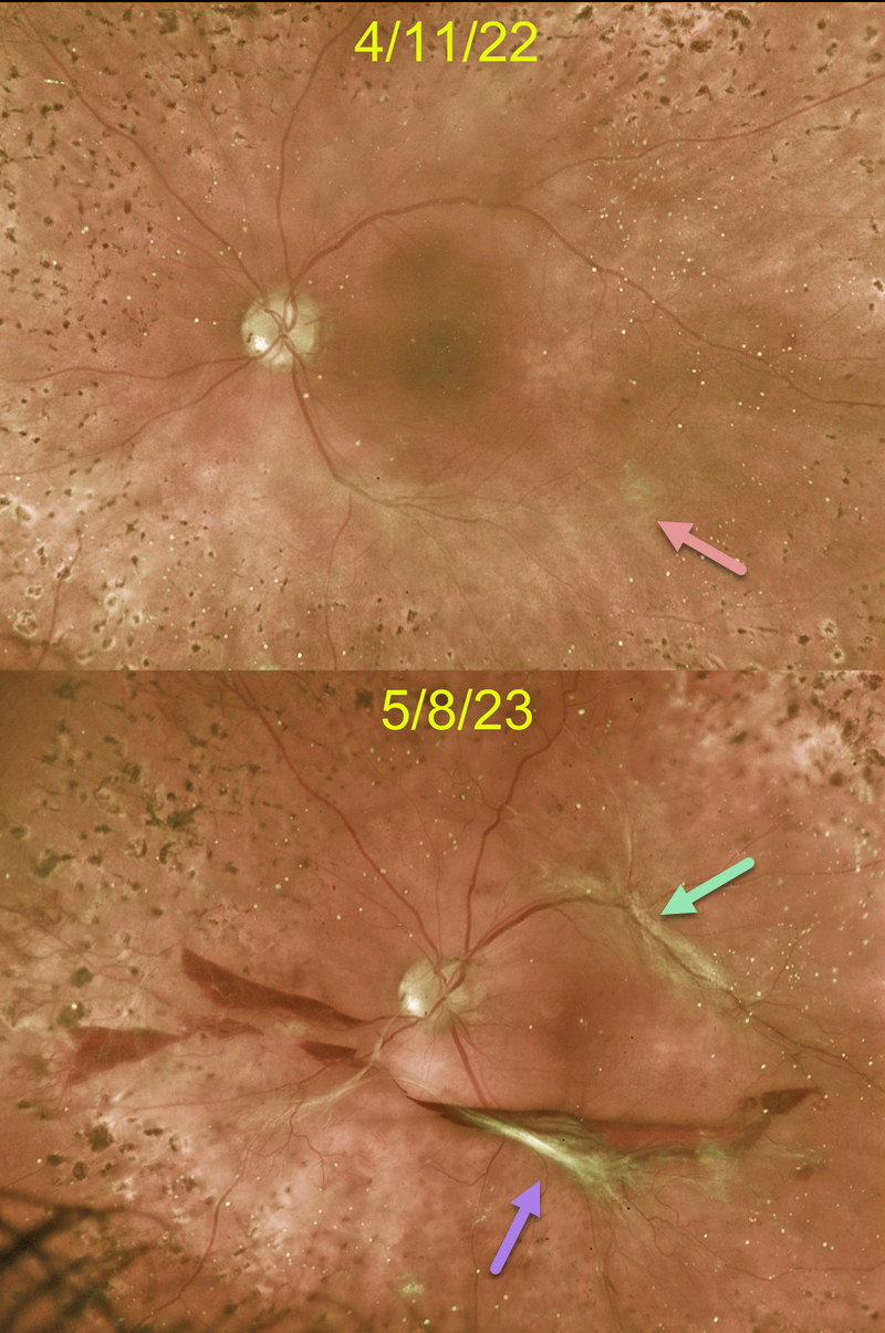
This 45YO female with type 1 diabetes responded beautifully to full panretinal photocoagulation (PRP) for her proliferative diabetic retinopathy (PDR). When examined 6 weeks following completion of her PRP, faint fibrotic neovascularization was noted along the inferotemporal arcade. Vision was 20/400 from residual central edema that was being treated with ongoing anti-VEGF therapy.
Unfortunately, she was lost to follow-up for over a year. Although vision remained at 20/400, increased traction was noted inferiorly, with new traction superiorly, resulting in an extrafoveal traction detachment. Symptomatic vitreous hemorrhage was also seen. Vitrectomy surgery was scheduled.
Learning Points:
Although PRP is often a ‘1 and done’ procedure, this is not always the case (Gonzalez et al, Ophthalmology 2021;128:1448-1457), and patients therefore need continued monitoring.
Although our patient showed involution of her PDR following PRP, increased fibrosis and traction still developed about a year later.
These findings would likely have been caught earlier if she had not been lost to follow-up, which is unfortunately quite common among patients with diabetes (Obeid et al., Ophthalmology 2018;125:1386-1392; Gao et al., Ophthalmology Retina 2019;3:230-236; Green et al, AJO 2020;216:18-27, etc.).



