

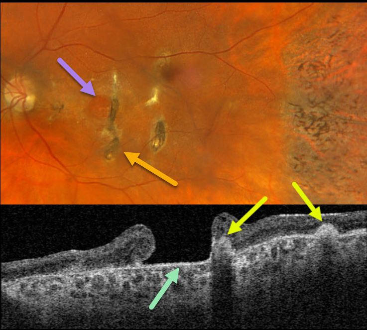
This 57YO female has a long history of vision loss in her left eye following blunt trauma years earlier. Vision was hand motion OS.
Optos color RGB imaging shows a large chronic macular hole with surrounding pigmentary changes. There are several hyperpigmented choroidal ruptures just temporal to the hole. Chorioretinal scarring with intraretinal pigment migration is noted in the temporal periphery.
Swept-source OCT shows a large macular hole. The choroidal ruptures appear as elevated hyperreflective subretinal mounds with posterior shadowing.
Learning Points:
This patient illustrates several sequelae of blunt trauma, including macular hole formation, choroidal rupture, and intraretinal pigment migration with a pseudo-sector retinitis pigmentosa appearance due to commotio-induced photoreceptor loss.
Since fresh traumatic holes can spontaneously flatten, especially in pediatric cases (Liu et al, AJO 2020;210:174-183), an initial period of observation is recommended before considering vitrectomy.
Given the chronicity of our patient’s hole, surgery was not considered.



