

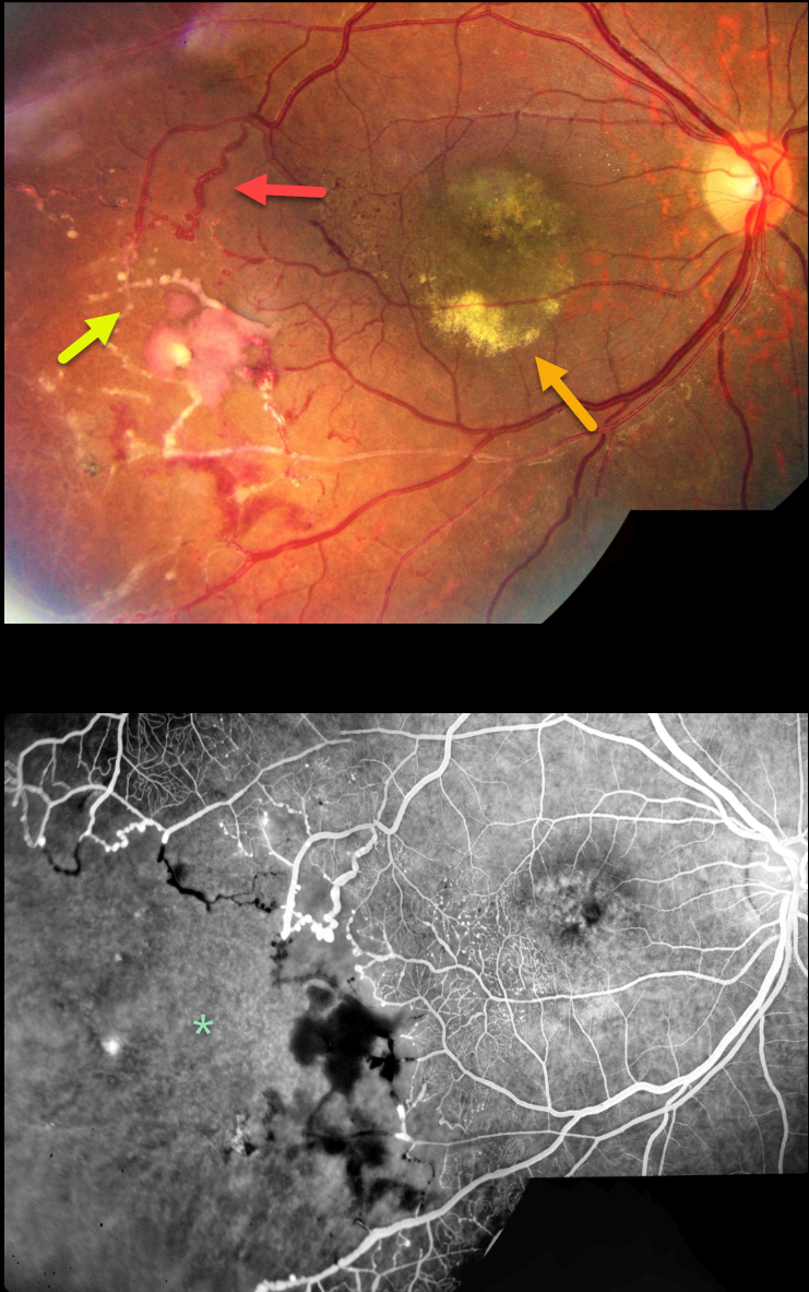
This 19YO female presented with a chronic history of unilateral vision loss in her right eye. Vision was 20/100 OD and 20/20 in her healthy left eye.
Color imaging shows extensive vascular changes in the inferotemporal retina, including retinal telangiectasia, macroaneurysms, vascular beading, sheathed vessels, and featureless ischemic retina. The central macula is hyperpigmented with variable subretinal lipid.
Fluorescein angiography shows marked distal capillary loss. The vessels bordering the perfused and ischemic retina are irregularly dilated with anastomoses. Fine telangiectatic vessels are noted in the inferotemporal macula.
Learning Points:
Coats disease is characterized by vascular changes throughout the retinal vasculature, including the arteries, the capillary bed, and the veins. Larger dilated vessels in Coats can sometimes mimic angiomas, vascular tumors, or exudation from retinoblastoma. The presence of retinal telangiectasia can help establish the diagnosis of Coats in these cases.
Coats are usually unilateral in males. However, recent ultra-widefield angiographic studies indicate subtle vascular changes mostly in the temporal periphery of the fellow eye (Brockmann et al, BJO 2021;105:1444-1453).
Dyskeratosis congenita or familial exudative vitreoretinopathy (FEVR) should be considered for patients with frank bilateral disease (Teixeira et al, Arch Ophthalmol 2008;126:134-135), in women with Coats-like findings (Johnson et al, Genetics 2009;30:181-184), or in patients with a family history of similar findings.
Laser photocoagulation can be used to directly treat leaking telangiectatic vessels, and a scatter laser can be placed into the ischemic retina. Anti-VEGF injections can also be used as primary or secondary treatment.



