

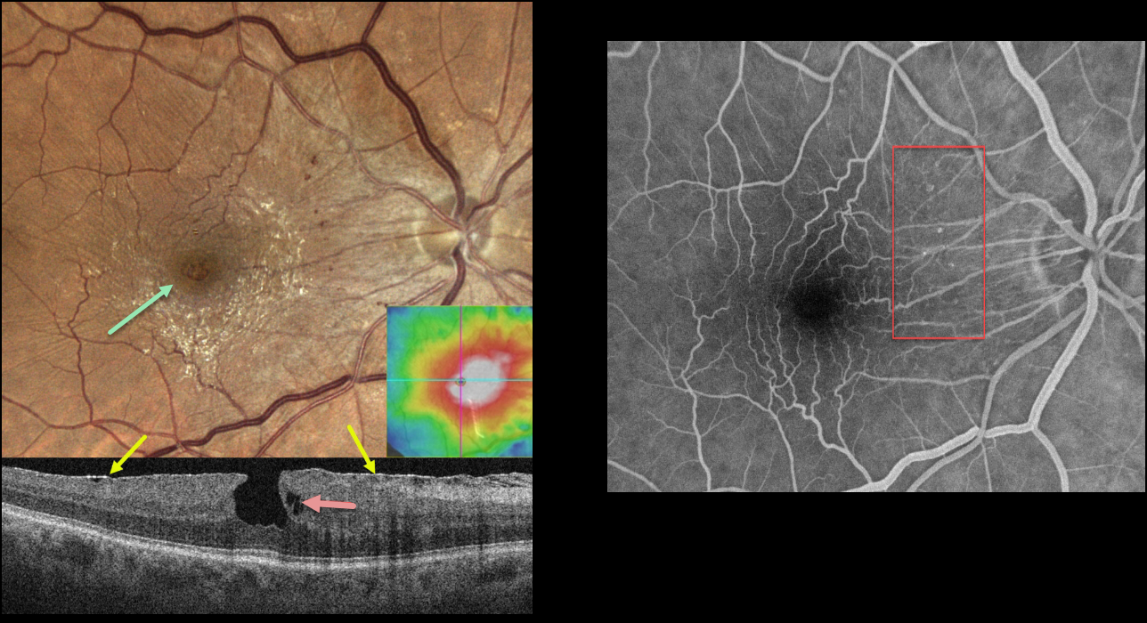
This 79YO male was referred for asymptomatic macular findings in his right eye. Vision was 20/20.
Color imaging shows a shiny, mostly nasal and inferior, macular pucker with a small, irregular macular pseudohole. Small dots of retinal blood vs telangiectasia are seen nasally.
OCT scanning shows an adherent macular pucker, a vertical foveal contour, and a small area of outer plexiform layer (OPL) schisis. The macula was diffusely thickened.
Fluorescein angiography shows no macular leakage, with filling of the nasal telangiectatic vessels.
Learning Points:
This case nicely illustrates many clinical and multimodal imaging findings for macular pucker. Clinically, true macular holes are virtually always perfectly round, whereas these pseudoholes are usually somewhat oval. The adherent epimacular tissue usually spares the foveal center. The adherent surrounding tissue contracts, drawing the surrounding tissue towards the macular center, giving the fovea a more vertical contour.
The Müller cell spans nearly the full thickness of the retina and helps mechanically hold the 3 layers of retinal cells together. Henle’s layer (the macular OPL) is inherently weakened since the photoreceptor axons and Muller cells are splayed laterally. Vitreomacular traction or traction from an overlying epimacular membrane will often therefore split Henle’s causing non-exudative schisis or cysts.
Finally, the secondary telangiectatic vascular changes are caused by mechanical damage from the overlying membrane. In severe cases, a pucker with the vascular changes can simulate a combined hamartoma.



