

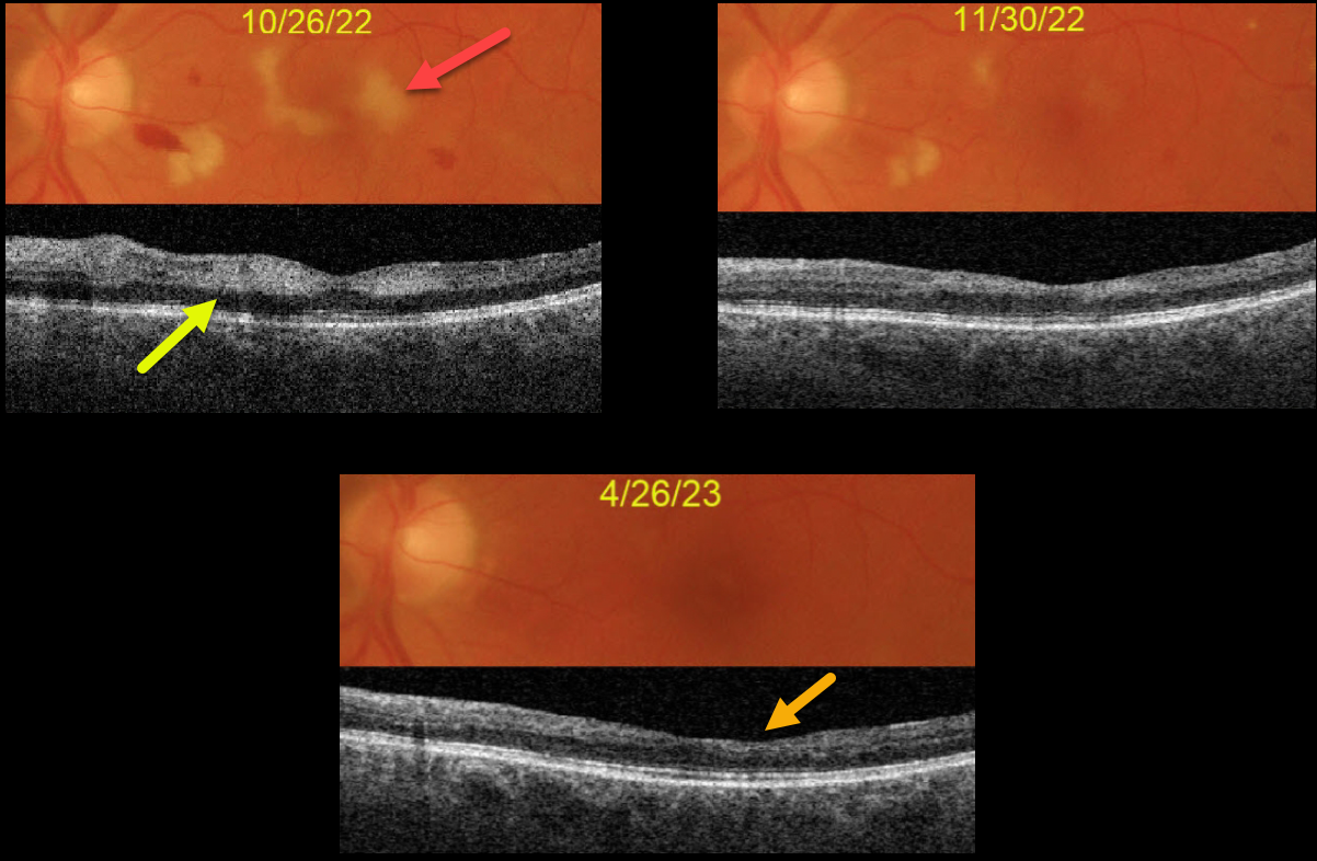
This 63YO male complained of vision loss in his left eye following left carotid endarterectomy. His blood pressure was markedly elevated perioperatively, but he reported feeling better. Blood pressure was 175/101 when seen by his doctor one week earlier. Vision was 20/25 OD and 20/200 OS.
Hypertensive retinopathy was noted bilaterally, with scattered retinal hemorrhages and nerve fiber layer infarcts (cotton-wool spots, CWS).
Color imaging of the left eye shows CWS virtually involving the macular center. Swept source OCT shows inner retinal hyperreflectivity from these lesions.
Over the following months, both his blood pressure and macular appearance normalized. When last seen on 4/26/23, vision was 20/40, although he was still quite symptomatic from paracentral scotomas due to inner retinal atrophy from the resolved CWS.



