

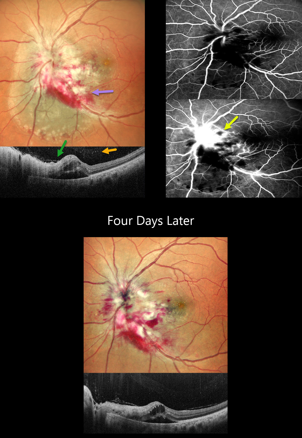
This 42YO female with a known history of HIV infection presented with 1 month of vision loss in her left eye. Her last CD4 count was 38 despite being compliant with HAART therapy. Her husband was HIV positive. Vision was 20/20 in her normal OD and 20/400 in OS.
Multicolor SLO imaging shows active cytomegalovirus (CMV) retinitis involving the nerve, which extends nasally and inferotemporally. Subretinal fluid is noted clinically and on OCT. OCT also shows full-thickness disorganization and hyperreflectivity, along with overlying vitreous cells. Fluorescein angiography shows early blockage with late disc and retinal leakage.
Oral valganciclovir and biweekly intravitreal injections were started. Four days later, vision improved to 20/90 with improvement in the papilledema and retinitis.
Learning Points:
CMV retinitis develops as a reactivation of latent CMV in immunosuppressed individuals. Before effective antiretroviral treatment emerged in the mid to late 1990’s, CMV retinitis developed in up to 40% of HIV/AIDS patients, often within the last 6 months of life. Treatment includes a combination of systemic and intravitreal medications, including ganciclovir, foscarnet, and cidofovir. Rhegmatogenous retinal detachment is a common late sequela and requires vitrectomy with silicone oil.



