

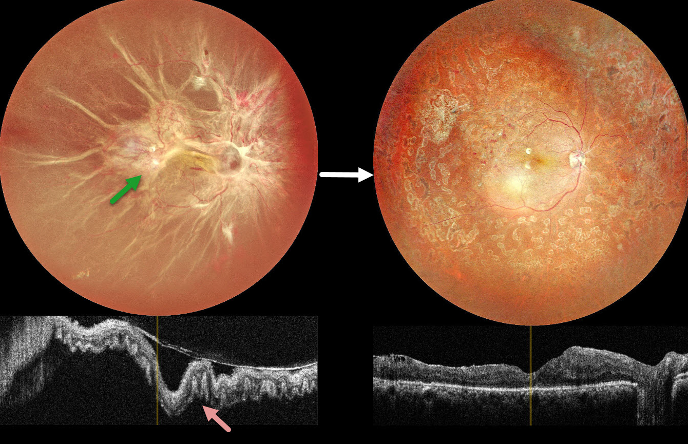
This 40YO female presented with vision loss in her right eye. There was a 15-year history of type 1 diabetes. Vision was hand motion OD.
Color photography shows a total combined traction-rhegmatogenous retinal detachment (RRD). There is a complex pattern of fibrovascular tissue covering the posterior pole with radiating retinal folds extending peripherally. OCT scanning shows a variably undulated detached retina with outer retinal corrugations.
Five days following an intravitreal anti-VEGF injection, she underwent pars plana vitrectomy with membrane peeling, endolaser, and silicone oil injection. Three months later, the silicone oil was removed. Postoperatively, vision improved to 20/80 with complete retinal reattachment.
Learning Points:
Outer retinal corrugations, also known as hydration outer retinal folds, are one of the key findings that help differentiate a RRD from exudative and traction retinal detachments. The cause of these folds remains uncertain but may result from the hydration of glycosaminoglycans in the interphotoreceptor matrix (Dalvin et al, Retinal Cases & Brief Reports 2020;14:355-359). This OCT biomarker confirms the rhegmatogenous component of our patient’s detachment.



