

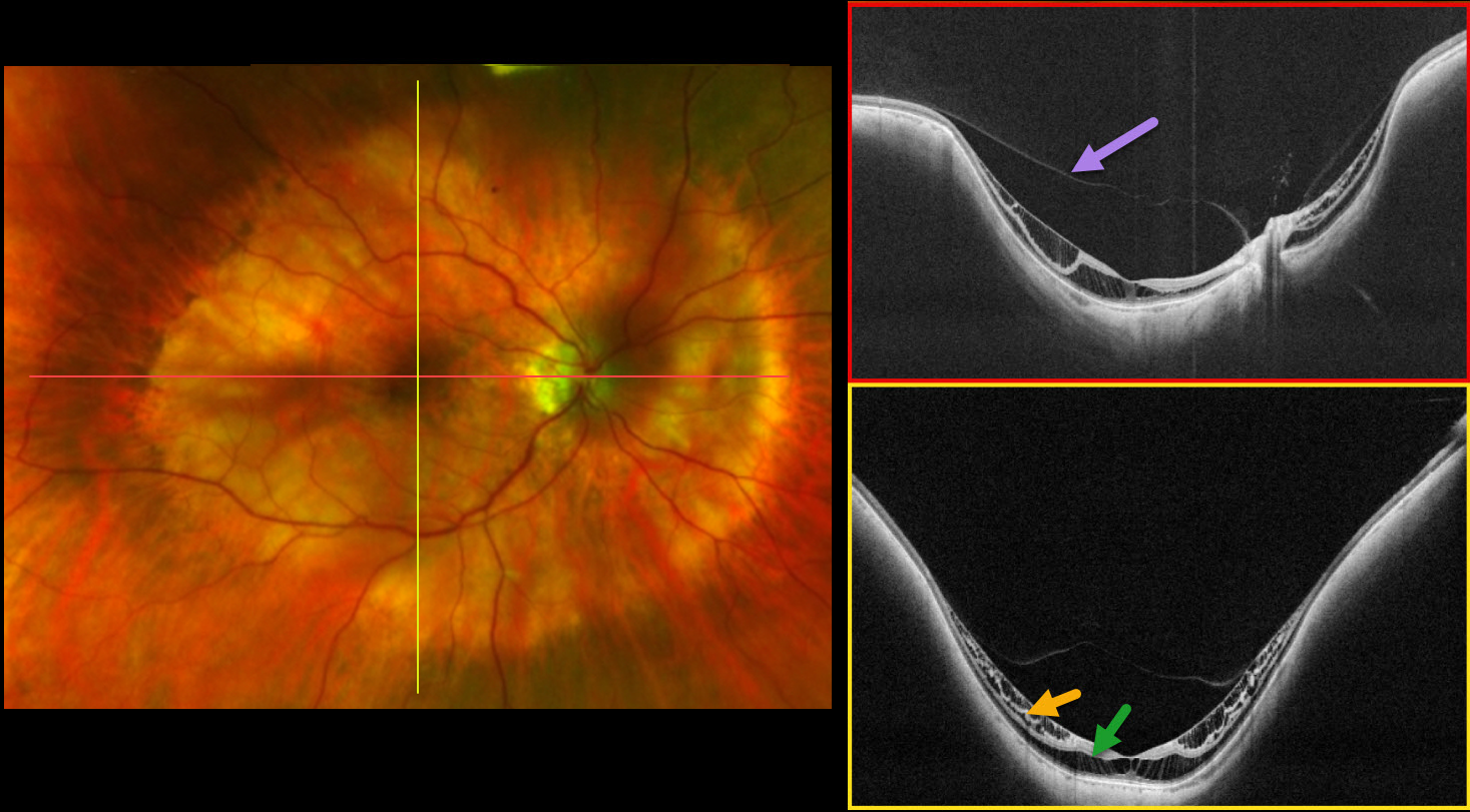
This 64YO female has a history of high myopia with posterior staphyloma. The axial length is 24.8 mm. Vision in the right eye has been deteriorating to 20/40.
Optos color RG imaging shows a posterior staphyloma, Type I (wide macula), in the right eye, which encircles the macula and disc. Ultra-widefield OCT (DreamOCT Intalight, 26 mm scan) shows a partial posterior vitreous detachment over the macula into the staphyloma with inner and outer retinal schisis within the staphyloma. There is no retinal detachment.
Learning Points:
Pathologic or high myopia is usually defined as a refractive error of at least -6 diopters with an axial length of at least 26.5 mm, so our patient technically is not a ‘high’ myope. 3D MRI imaging allows for a more nuanced definition and has shown that the ocular deformations can affect the entire eye, the equatorial, or just the posterior globe (Luo et al, JAMA Ophthalmology 2023;141:768-774). As defined by Luo et al, our patient has a posterior bulb-shaped globe. The ultra-widefield OCT is designed to image the entire posterior profile of the eye, which shows the geometry of our patient’s staphyloma.
The image also shows that myopic traction maculopathy (MTM) underlies a partial vitreous detachment. This confirms that the main cause of MTM is not the vitreous but the posterior staphyloma. Furthermore, when looking at the horizontal scan, the thickness of the retina (or, in other words, the severity of the schisis) is not evenly distributed into the staphyloma. It is more pronounced in the temporal side of the macula. The optic nerve pushes the sclera anteriorly, buckling the retina in that area and releasing the traction.
In the vertical scan, the schisis is uniformly distributed in the whole staphyloma. According to the ATN (atrophic, tractional, and neovascular) classification system for myopic maculopathy (Ruiz-Medrano et al, Progress Retinal Eye Research 2019;69:80-115), our patient has A1 (tessellated fundus only), T2 (inner + outer foveoschisis), and N0 (no myopic CNV). According to the myopic traction maculopathy staging system (MSS, Parolini et al, Ophthalmic Surg Lasers Imaging Retina 2023;54:153-157), our patient is affected by MTM in Stage 2a (predominantly outer macular schisis with no macular hole).



