

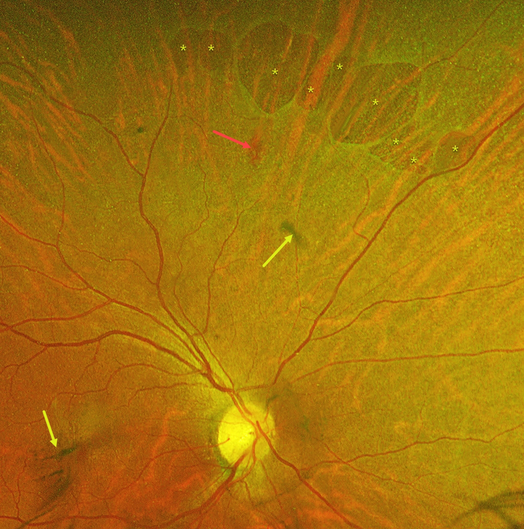
This 55YO female was referred with mildly decreased vision in her right eye two days following blunt ocular trauma. Vision was 20/40 OD and 20/20 in her normal OS.
Optos color RG imaging reveals numerous irregularly oval superonasal retinal holes, a retinal hemorrhage, and some clumps of vitreous blood. There was no vitreous detachment clinically.
Learning Points:
Retinal breaks following blunt trauma include retinal dialysis, horseshoe tears, and giant retinal tears. Breaks can also develop from concussive injury at the impact site or from a coup-contrecoup injury, so-called impact or necrotic breaks, which may be associated with varying degrees of commotio retinae acutely (Cox et al, Arch Ophthalmol 1966;76:678-685) or following its resolution (Longstaff et al, Br J Ophthalmol 1987;71:375-376).
In this case, the breaks identified 2 days after the injury were not associated with commotio. However, their appearance and the adjacent hemorrhage support an acute traumatic etiology. Therefore, prophylactic laser retinopexy was promptly applied around all breaks.



