

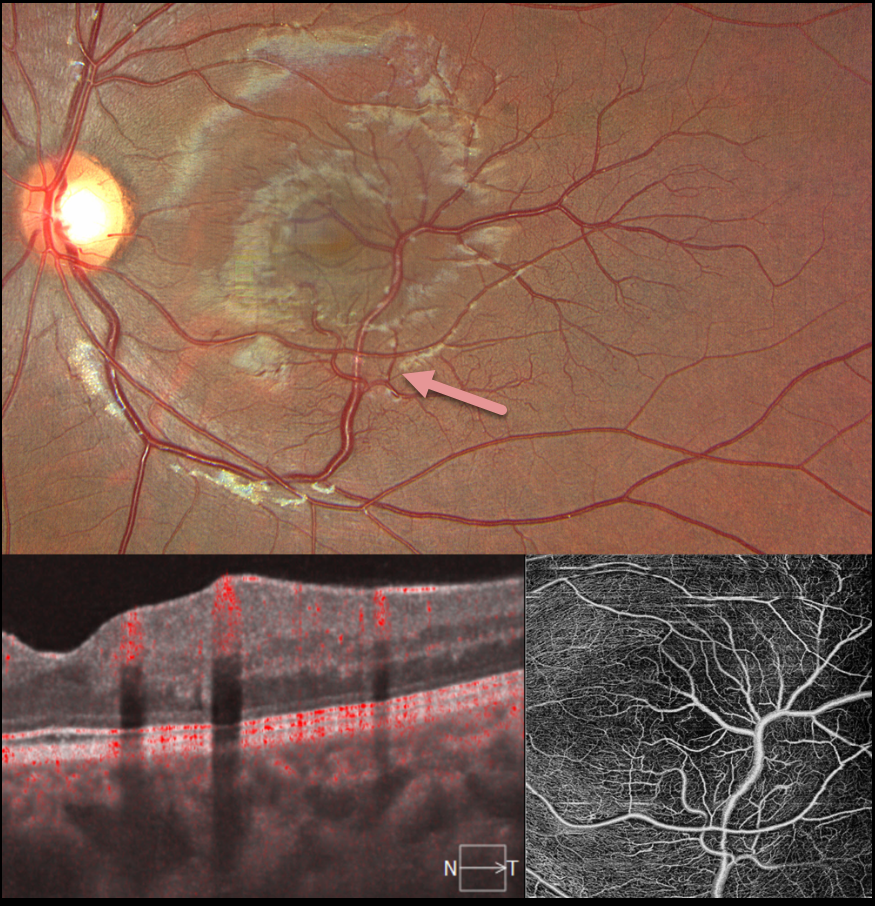
This 17YO female presented for a routine eye examination. Vision was 20/20 in her normal OD and 20/20 OS. Color imaging shows an inferior retinal venous malformation that drains the inferior and superotemporal macula. En face and B-scan OCT angiography show these vessels to lie in the inner retina.
Learning Points:
Retinal vessels virtually always respect the horizontal meridian. Retinal venous malformations are congenital anomalous vessels that, by definition, cross the horizontal. Originally described by Gary Brown et al as congenital retinal macrovessels (Arch Ophthalmology 1982;100:1430-1436), these vessels are usually located in the macular region and are virtually always venous. These lesions are now called retinal venous malformations, since about one-quarter of patients have associated venous anomalies in the brain (Pichi et al, JAMA Ophthalmol 2018;136:372-379).



