

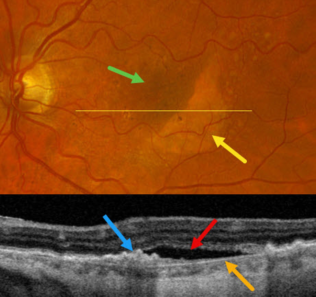
This 71YO female complained of one month of decreased vision in her left eye. Vision was 20/30 OD and 20/200 OS.
Moderate larger drusen with pigment clumps were noted in her right eye (not shown). Optos color RGB imaging of her left eye shows a crescent of subretinal pigment loss in the inferotemporal macula with increased subretinal pigment superonasally. OCT scanning within the area of pigment loss shows bare Bruch membrane with overlying subretinal fluid. The RPE nasally is retracted and bunched onto itself.
Learning Points:
A tear of the RPE is most seen in RPE detachments (PEDs) associated with neovascular AMD, but can also occur in numerous other etiologies, including central serous chorioretinopathy and polypoidal choroidal vasculopathy. The tear leaves a depigmented region where the RPE was and is now absent, along with a pigmented, relatively straight subretinal lesion from the scrolled RPE edge.
Our patient may harbor a macular neovascularization beneath the retracted RPE. A therapeutic trial of monthly intravitreal Avastin was thus started.



