

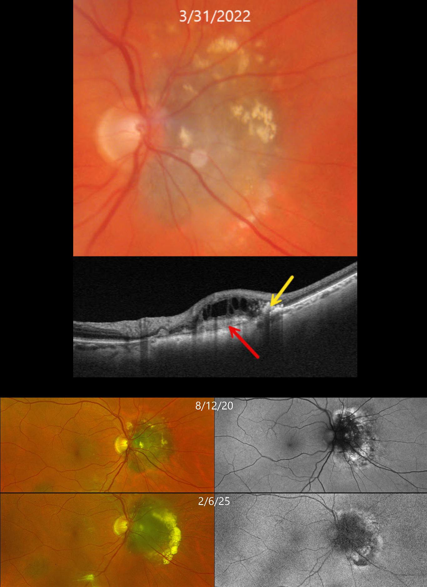
This 71YO female has been followed for several years with a choroidal nevus and chronic overlying choroidal neovascularization (CNV).
On March 31, 2022, Triton color photography shows a nasal peripapillary choroidal nevus that touches the optic nerve. Lipid is noted mostly along its superior and temporal edges. Swept-source OCT shows a hyperreflective type 2 (above the RPE) CNV, hyperreflective lipid, and overlying cystic retinal thickening.
The CNV has been observed due to her complete lack of symptoms and extramacular location of the lesion. When last examined on February 6, 2025, the nevus looks fairly stable on Optos RG imaging, although there is increased subretinal lipid along its temporal edge. On fundus autofluorescence (FAF), the CN is hypo-FAF with hyper-FAF along its periphery.
Learning Points:
Choroidal neovascularization is a rare finding in choroidal nevi, and associated exudation can be misinterpreted as exudation from possible malignant transformation. OCT angiography is helpful in diagnosing these lesions, which respond well to either intravitreal anti-VEGF monotherapy or combination treatment with photodynamic therapy (Munie and Demirci, Ophthalmology Retina 2018;2:53-58).



