

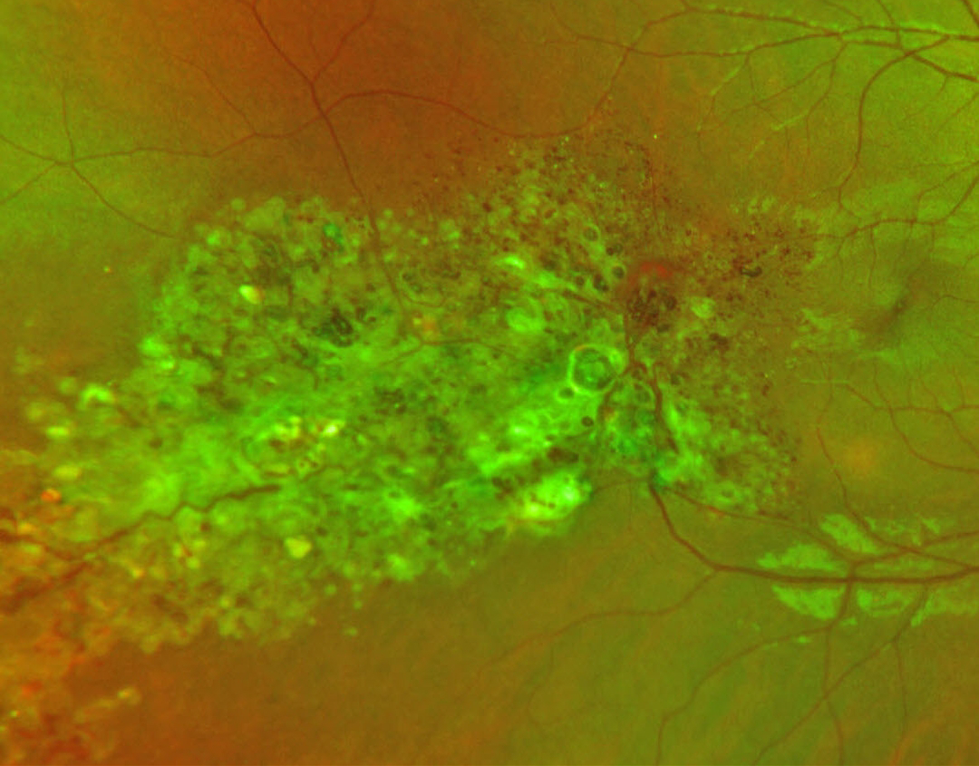
This healthy 25YO female presented with an asymptomatic retinal lesion in her right eye. She recalled being diagnosed with a retinal finding in the past and reported negative prior neurologic imaging. Vision was 20/20 bilaterally.
Optos RG color imaging shows extensive confluent variably sized grape-like clusters of aneurysms filled with dark venous blood. Some scattered gray fibrotic membranes cover its superficial surface.
Learning Points:
Retinal cavernous hemangiomas are usually unilateral, benign vascular abnormalities. Fluorescein angiography shows plasma-erythrocyte layering within these saccular lesions caused by sluggish blood flow. Vision is usually normal, and no treatment is required. Rarely, vision loss can occur from vitreous hemorrhage. Although most occur sporadically, some may be associated with an autosomal dominant neurocutaneous syndrome, including cutaneous and central nervous system hemangiomas. Neurologic imaging is therefore recommended.
Unfortunately, our patient was immediately lost to follow-up.



