

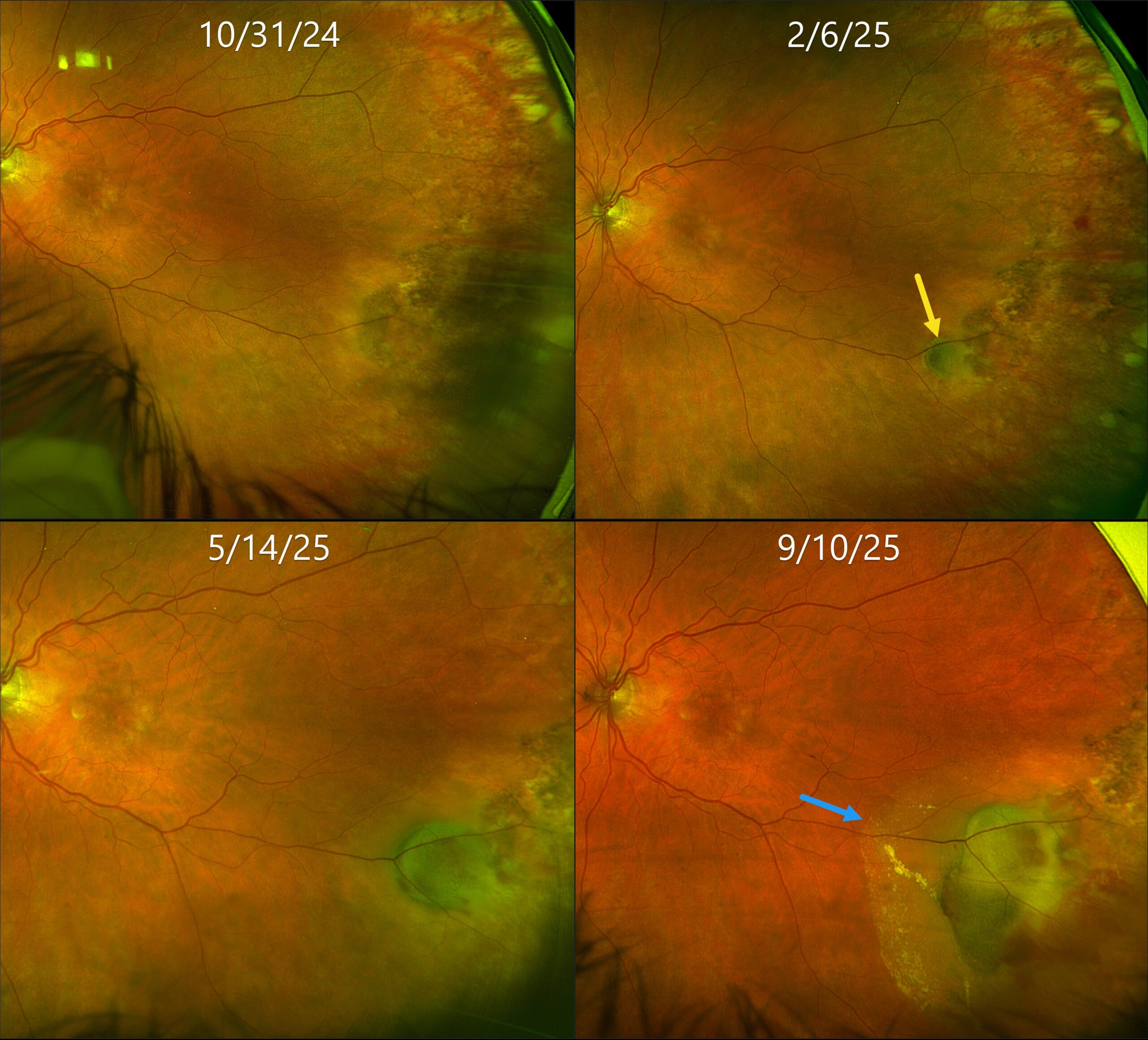
This 80YO female was initially referred for asymptomatic retinal findings in her left eye. Vision was 20/25.
Optos color RG imaging shows flat pigmented subretinal scarring in the left inferotemporal periphery. Three months later, a subretinal pigmented nodular lesion is noted along with a small dot of temporal blood. Over the following 7 months, this subretinal lesion enlarged with increasing blood and subretinal fluid approaching the macula. Although she remained asymptomatic, intravitreal anti-VEGF therapy was started.
Learning Points:
Ectopic choroidal neovascularization (CNV), commonly referred to as peripheral exudative hemorrhagic chorioretinopathy (PEHCR), is an exudative process similar to wet AMD but located in the retinal periphery. Ectopic CNV can often masquerade as a choroidal mass or uveal melanoma. It can also cause extensive choroidal/suprachoroidal, subretinal, and vitreous hemorrhage, more commonly in patients who are on blood thinners. The subretinal blood often spontaneously regresses without treatment, although anti-VEGF injections can be used for symptomatic or increasing exudation.



