

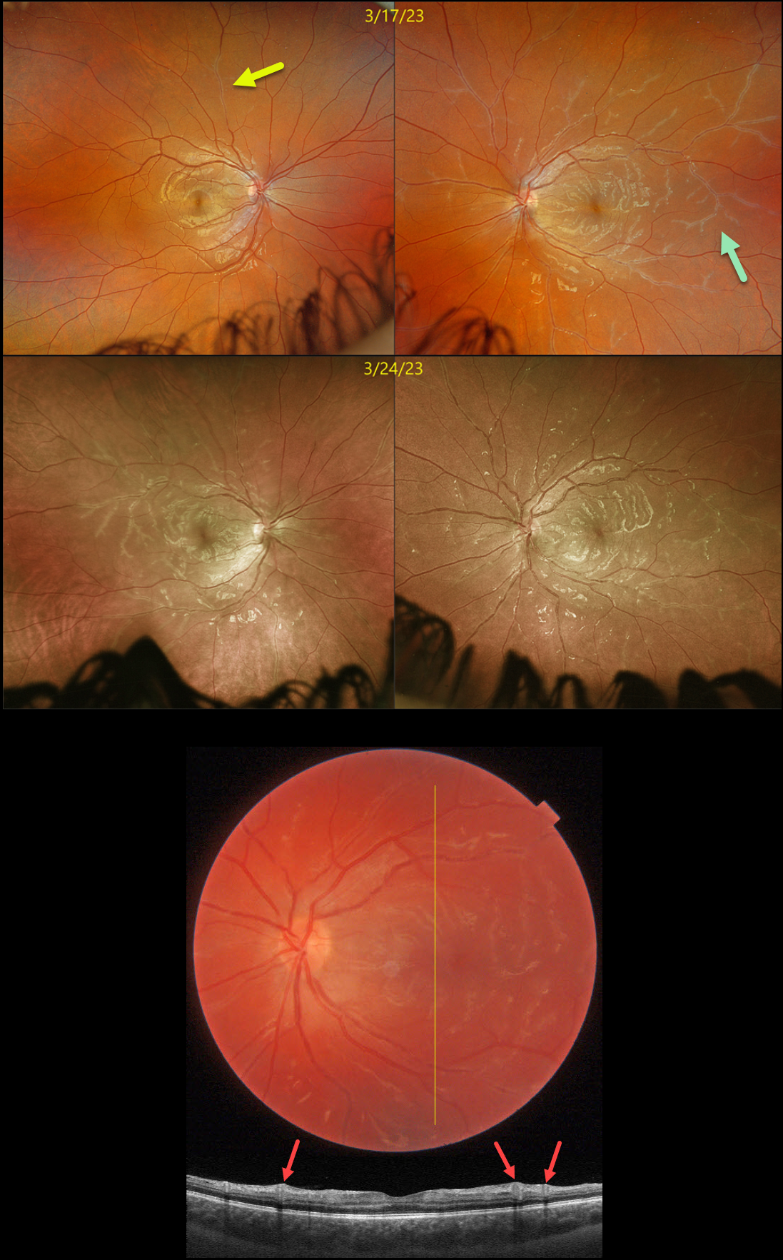
This healthy 16YO female presented with several days of bilateral blurred vision. There was no past ocular or medical history. Vision was 20/25 OD and 20/30 OS. Anterior segments were normal.
Optos imaging of her right eye shows a frosted branch appearance to a retinal vein in the superior midperiphery, with more diffuse vasculitis seen in her left eye.
Swept-source OCT through the left macula shows some hyperreflective inner retinal thickening around the inflamed veins. Bloodwork for Bartonella, syphilis, and sarcoid was ordered.
She returned one week later with new blurred vision in her left eye. Vision was 20/25 OU. Although findings were stable OS, new diffuse frosted branch changes were present in the OD. Her bloodwork returned negative, and she was started on oral prednisone 60mg daily.
Learning Points:
Frosted branch angiitis is a retinal vasculitis that gets its name from the involved vessels resembling branches of a tree that are “frosted” with snow (Kleiner et al, AJO 1988;106:27-34).
Although these findings may be isolated, they are often considered a phenotype found in a multitude of inflammatory conditions, most commonly CMV retinitis. The inflammation is usually very responsive to steroids, and the visual prognosis is generally good, although some patients may develop secondary anterior and posterior segment neovascularization due to widespread ischemia.



