

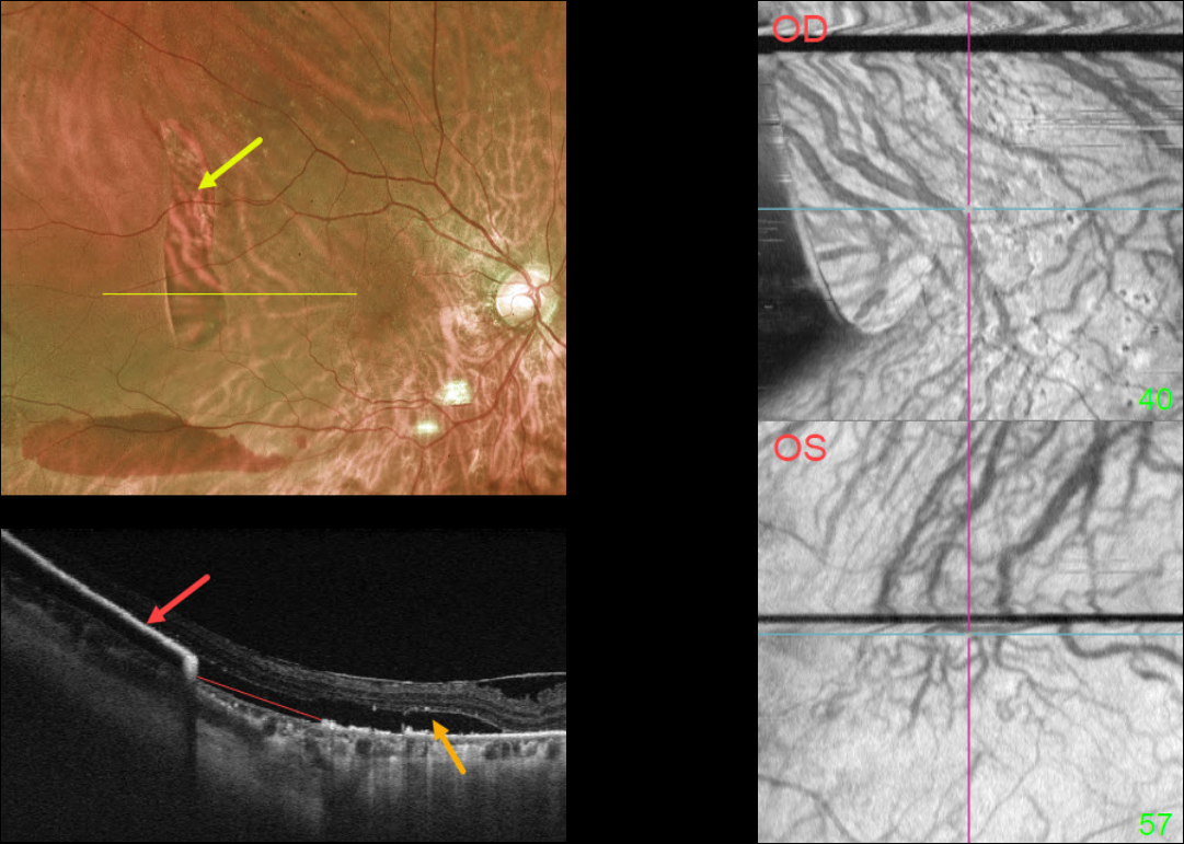
This 73YO female presented with acute vision loss in her right eye. Vision was 20/40 OD and 20/30 OS. There was a several-year history of type 2 diabetes with mild bilateral nonproliferative diabetic retinopathy.
Optos imaging shows a large vertical area of depigmentation in the distal superotemporal macula. There is a serous detachment surrounding this region, along with a far inferior subretinal hemorrhage.
Swept-source OCT through this lesion shows bare Bruch’s membrane from an RPE tear. The retracted RPE is markedly hyperreflective more temporally. Subretinal fluid overlies this region.
Macular choroidal en face imaging shows bilateral anomalous vasculature with choroidal anastomoses.
Learning Points:
A tear of the RPE is most commonly seen in RPE detachments (PEDs) associated with neovascular AMD, but can also occur in numerous other etiologies, including central serous chorioretinopathy and polypoidal choroidal vasculopathy.
The tear leaves a depigmented region where the RPE was and is now absent, along with a pigmented, relatively straight subretinal lesion from the scrolled RPE edge.
The choroidal en face OCT findings are more consistent with the pachychoroid spectrum. RPE tears are found in polypoidal choroidal vasculopathy, another pachychoroid disorder (Zhao et al, Retina 2020;40:477-489).
See Ersoz et al for a great review on RPE tear classification, pathogenesis, predictors, and management (Surv Ophthalmology 2017;62:493-505).



