

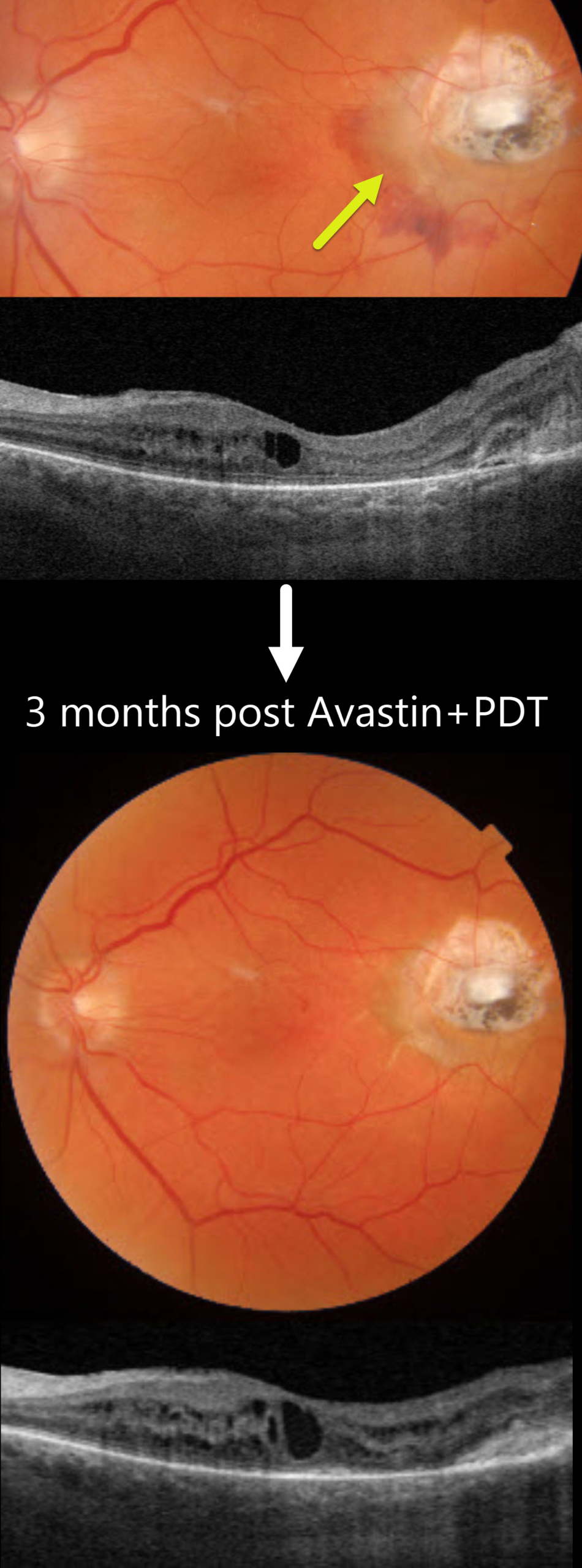
This 75YO male underwent successful vitrectomy for a macula-off rhegmatogenous retinal detachment (RRD). Vision improved from a preoperative hand motion vision to counting vision 7 months following surgery. However, he returned 4 months later with new distortion and blurriness. Vision was 20/400.
Triton color imaging shows an active choroidal neovascularization (CNV) with associated subretinal blood along the temporal and inferior edge of the drainage retinotomy scar. Swept-source OCT shows mild macular cystic edema arising from the temporal type 2 (above the RPE) CNV. The peripheral retina remained attached.
The lesion failed to respond to several intravitreal Avastin injections, necessitating combined Avastin and photodynamic therapy. Exudation completely resolved 3 months following this treatment.
Learning Points:
Invented by Steve Charles in the early days of vitrectomy, the drainage retinotomy has become an integral part of retinal reattachment surgery (Ramamurthy et al, Surv Ophthalmology 2023;68:1038-1049). Following the adage that ‘every time a hole is made in the retina, some vision leaks out,’ it is always preferable to drain subretinal fluid through a pre-existing break whenever possible. When required, the site should ideally be located superiorly and well away from the macula. Drainage retinotomies are associated with an increased risk of macular pucker and cystoid macular edema (McKay et al, AJO 2022;241:227-237). CNV is an exceedingly rare complication, which may be caused by inadvertent RPE-choroidal touch by the drainage needle or thermal-induced damage.



