

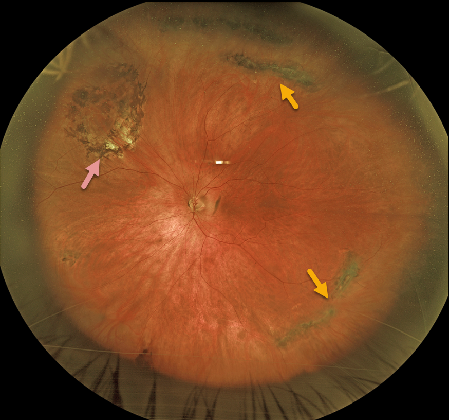
This 68YO male presented with asymptomatic lattice degeneration bilaterally. He remembered getting a laser to one of his eyes many years earlier at an outside practice.
Optos imaging of his left eye shows a near ora to ora view of scattered circumferential pigmented lattice lesions. Laser scarring surrounds an area of lattice with atrophic holes superonasally.
Learning Points:
Lattice lesions have a pocket of vitreous liquefaction overlying the thinned retina with a firm vitreo-retinal adhesion along their borders. White, fibrosed vessels often criss-cross the lesions, which leads to the name, lattice. This firm vitreoretinal adhesion, along with the often-present atrophic holes, can predispose these eyes to retinal detachment.
Retinal detachment occurs in 1 in 10,000 people per year. Lattice is found in 10% of the general population, especially in high myopes.
Since about one-third of patients with retinal detachment have lattice as the cause, the question always arises, especially in patients like this: Should prophylactic retinopexy be performed? The short answer is that asymptomatic lattices generally should not be treated.
To put things into perspective, imagine a sports stadium of 30,000 people. Three people will develop a retinal detachment in the coming year. Only one of the 3,000 people in the stadium with lattice would develop one of those 3 detachments. Assuming prophylactic treatment is 100% effective with no complications, 2,999 eyes would be unnecessarily treated to prevent one detachment.



