

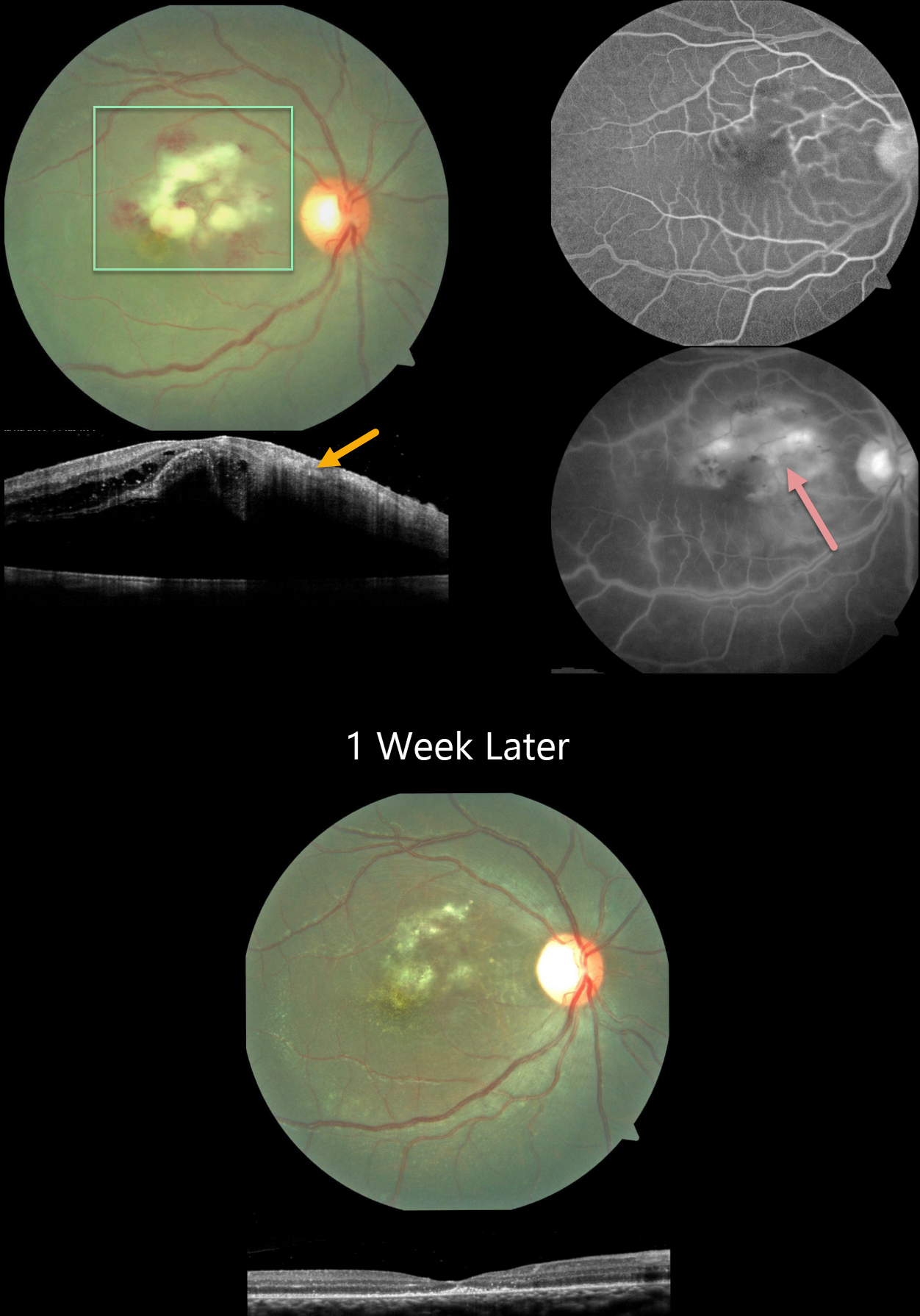
This 15YO boy had a 3-week history of Dengue fever followed by severe vision loss in his right eye. Vision was counting fingers OD and 20/20 in his normal left eye.
Color imaging shows Purtscher-like central macular findings of acute inner macular ischemic whitening with retinal hemorrhages. OCT scanning shows retinal thickening with increased inner retinal hyperreflectivity. Fluorescein angiography shows leakage and staining from the ischemic tissue.
Dengue testing was positive for IgG and negative for IgM antibodies. Weil Felix testing was positive for OX2 and OX19, indicating a likely rickettsial coinfection. Oral antibiotics and steroids were started.
One week later, vision improved to 20/120. The macular findings were resolving. OCT scanning shows complete resolution of the prior edema with secondary thinning and disorganization of the outer foveal layers.
Learning Points:
Epidemic (post-fever) retinitis was described by Kawali et al (Ocular Immunology & Inflammation 2019;27:571-577) as an infectious or para-infectious uveitis occurring days to weeks following a febrile illness due to Dengue, Rickettsiosis, West Nile, Leptospirosis, Chikungunya, Typhoid, and Ebola. It appears as a usually bilateral Purtscher ’s-like retinitis involving the posterior pole with a favorable response to steroids. It is most common in tropical countries like India. See Mahendradas et al (Indian J Ophthalmol 2020;68:1775-1786) for an excellent post-fever retinitis review.



