

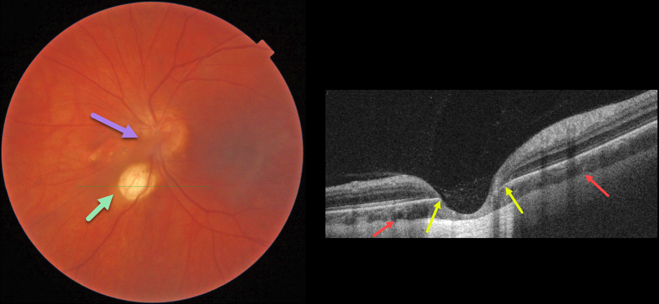
This 35YO female has a history of decreased vision in her left eye since childhood. Vision was 20/20 in her right eye and stable at 20/200 in her left eye. Anterior segments were normal except for a posterior polar cataract OS.
Color imaging of the left posterior pole shows a fairly round, one-disc-diameter depigmented lesion just inferonasal to the disc. An area of non-specific pigmentary changes is noted just nasal to the nerve, along with persistent fetal vasculature (hyaloid artery).
OCT through the depigmented lesion shows a choroidal coloboma. The underlying sclera is intact and slightly bowed posteriorly. Within the coloboma, there is loss of the outer retinal layers with draping of the inner retina against the bare sclera. The RPE is also absent within the coloboma.
Learning Points:
Choroidal colobomas result from a failure of closure of the optic vesicle and choroidal fissure during normal fetal development, but can also be caused by congenital toxoplasmosis and Zika virus. They can be solitary or multiple, and the visual potential depends on the extent of optic nerve and macular involvement. Patients with choroidal colobomas are at an increased risk for retinal detachment and macular neovascularization.
Embryologically, the hyaloid artery is vital to the proper development of the anterior segment, and usually regresses at about 10 weeks of gestation; however, it may persist in some cases. In severe cases, the eye may be microphthalmic with leukocoria and retinal traction. These eyes are typically amblyopic. A genetic association has not been identified.



