

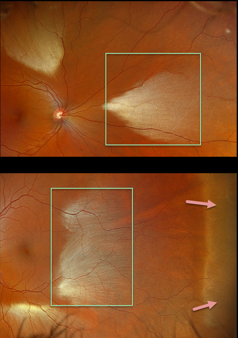
This healthy 35YO male was referred for asymptomatic fundoscopic changes. Vision was 20/20 OU.
Opto imaging shows large multifocal patches of myelinated nerve fiber layer (NFL) in both eyes. Temporal white without pressure (WWP) is also noted in his left eye.
Learning Points:
Myelinated NFL is a relatively uncommon, unilateral or bilateral, usually isolated finding present at birth. The myelinated NFL is often connected to the optic nerve, but not necessarily as in this case. Most patients are asymptomatic, although some may have a corresponding relative scotoma. The myelination can progress in about 10% of cases and can also resolve following retinal ischemic events.
Myelinated NFL occurs when retinal nerve fibers develop a myelin sheath, which usually stops posterior to the lamina cribrosa. The myelination may represent an oligodendrocytic choristoma (see Rao et al Retina 2019;39:1125-1132).
WWP is a relatively common benign peripheral finding, most often seen in darkly pigmented individuals. Typically, there are bilateral peripheral geographic areas of whitish retinal discoloration. WWP gets its name because the area appears white without indentation, unlike scleral depression (white with pressure). It can sometimes be confused with retinal breaks or detachment.
The exact cause of this phenomenon is unknown. Historically, it was felt to be due to a benign vitreoretinal interface change, but more recent OCT findings show increased reflectivity in the outer retina (see Diaz et al, Retina 2014;34:1020-1021).



