

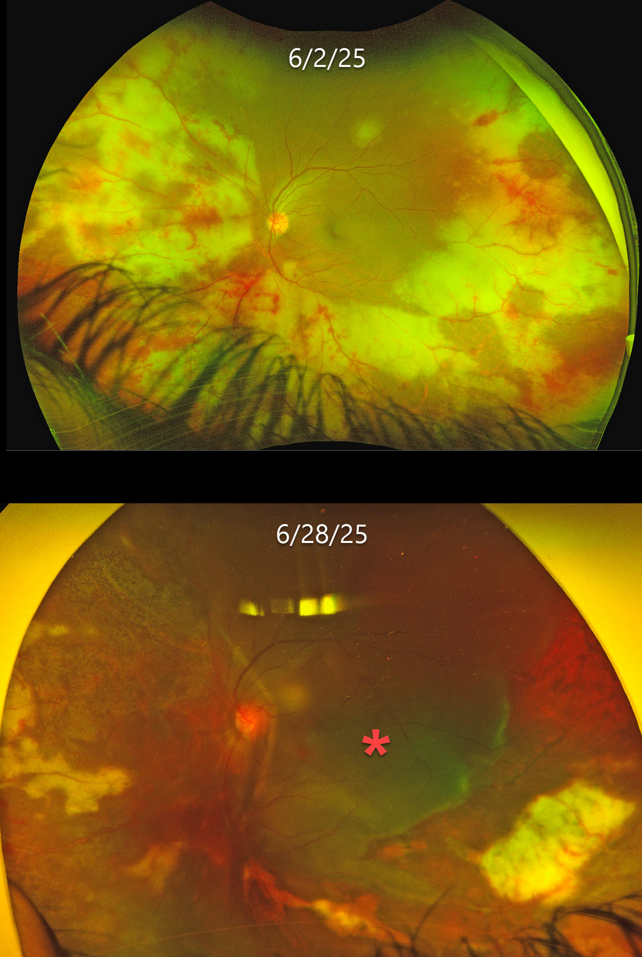
This previously healthy 35YO male presented with 1 week of floaters in his left eye. Vision was 20/20 in his normal OD and 20/20 OS. Anterior segments were normal.
Optos color RG imaging shows variably confluent outer retinal opacification with retinal hemorrhages extending from the periphery into the posterior pole. The retinal vessels are variably narrowed. He was initially diagnosed with possible cytomegalovirus (CMV) retinitis, started on valganciclovir, and referred to infectious diseases (ID).
He returned 3 weeks later with counting fingers vision despite improved retinitis and stopping systemic treatment after 10 days due to financial limitations. He returned 5 days later with sudden loss of vision. Vision was light perception with improved retinitis but with a new macula-off retinal detachment, which was repaired with lensectomy, vitrectomy, scleral buckle, retinectomy, and silicone oil. Two months later, vision was 20/150, and the retina remained completely attached.
Learning Points:
Acute retinal necrosis syndrome (ARNS) is usually caused by the herpes zoster virus. It generally occurs in immunocompetent individuals and presents as rapidly progressive panuveitis with hemorrhagic and ischemic unilateral (ARNS) or bilateral (BARNS) retinal necrosis, typically beginning in the retinal periphery.
Our patient was eventually evaluated by ID, who diagnosed him with HIV and a CD4 count of 45 with initiation of HARRT therapy. It is unclear if our patient’s underlying previously undiagnosed HIV infection was contributory to his retinitis. The lack of the usual anterior chamber and vitreous inflammation is likely due to his immunosuppression.
Both CMV retinitis and ARNS cause retinal necrosis and hemorrhages. However, CMV usually starts in the posterior pole, often beginning in a paravascular location, which then spreads outwards. The retinitis involves the full-thickness retina, so the hemorrhages and opaque retina also involve the inner retina and obscure the underlying larger retinal vessels. Our patient’s findings are more pathognomonic for ARNS, given the outer retinal findings that likely began in the retinal periphery and extended posteriorly, with no paravascular location.
ARNS is often a devastating and blinding infection due to the high risk for retinal detachment as well as macular and optic nerve involvement. The latest meta-analysis argues for systemic antiviral therapy and prophylactic vitrectomy, with uncertain benefits for prophylactic laser photocoagulation or adjunctive intravitreal antivirals (Zhao et al, Retina 2021;41:965-978).
Treatment should be initiated as soon as the diagnosis is suspected, without waiting for laboratory results. Oral valacyclovir is clinically equivalent to intravenous acyclovir (Baltinas et al, AJO 2018;188:173-180), and aspirin is often recommended to minimize vascular thrombosis. Although valganciclovir is not the treatment of choice, our patient’s retinitis still rapidly improved despite only 10 days of therapy. Treatment is continued until all retinitis becomes inactive, and usually for at least 3-6 months thereafter to reduce the risk of fellow-eye involvement.



