

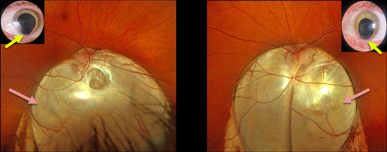
This 30YO female presented with a 6-month history of vision loss in her right eye. Despite being born with bilateral iris and choroidal colobomas, her vision in each eye was excellent until recently. Vision was 20/200 OD and 20/40 OS.
Optos RGB imaging shows bilateral inferior choroidal colobomas extending from just superior to the optic nerves towards the inferior peripheries. A shallow macula-off retinal detachment extends from the temporal edge of the right coloboma into the 7 to 10 o’clock periphery. In the office, we could not identify any retinal breaks. At vitrectomy, a small anterior retinal tear was found at the superior edge of the detachment.
Learning Points:
Choroidal colobomas are caused by failure of the optic vesicle and choroidal fissure to close during fetal development. Choroidal coloboma can present as solitary or multiple lesions, as in this case. Visual prognosis depends on the location and involvement of the optic nerve and macula.
The retina within the coloboma is variably thinned, increasing the risk of rhegmatogenous retinal detachment (RRD). More centrally within the coloboma, the retina consists of a monolayer of atrophic glial structures and neurons, known as the intercalary membrane (ICM). Between the edge of the coloboma and the ICM is a region of thinned retina lacking RPE and choroid, the marginal ICM (MICM). Breaks within the MICM, which extend about 2 disc diameters from the coloboma margin, are usually the cause of RRD (Tanaka et al, Ophthalmology Retina 2021;5;702-710).
Fortunately, our patient had a more typical RRD that was independent of her coloboma. Laser treatment along the edge of the coloboma was therefore not performed, thereby avoiding the risk of iatrogenic paracentral vision loss. The retina remained attached 5 weeks postoperatively, and vision improved to 20/100.



