

This 79YO female presented for a routine examination. ‘Successful’ macular hole surgery was performed elsewhere at least several years earlier.
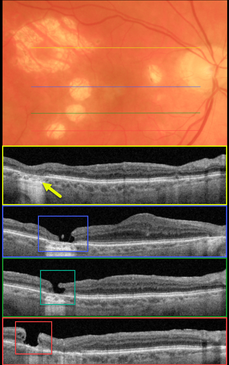
Optos color imaging shows multiple atrophic scars in the temporal macula with a smaller scar nasally. Swept-source OCT scanning through the most superior, larger scar shows outer retinal atrophy.
OCT scans through each of the 3 smaller temporal scars show independent macular holes. Scanning of the central macula shows a thickened retina just nasal to fixation.
According to our patient, the single vitrectomy was uneventful, but her imaging suggests otherwise. It seems the surgeon made at least 5 deep retinal touches with the intraocular forceps while attempting to peel the internal limiting membrane. The larger superotemporal scar was probably a touch that was surrounded by endolaser.
The 3 ectopic macular holes may have developed intra- or post-operatively. Fortunately, her central macular hole closed, her vision was 20/40, and she remains thrilled with her surgical results.



