

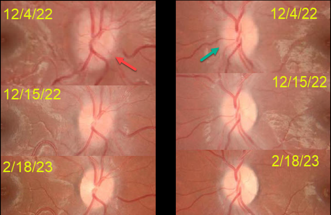
This 10YO boy presented on 12/4/22 with acute blurred vision in his right eye. He was hospitalized 2 weeks earlier for a viral illness with musculoskeletal pain and fever.
Vision was counting fingers OD and 20/20 OS. An afferent pupillary defect was noted OD. The right disc was moderately swollen, and milder disc swelling was noted OS. MRI was suggestive of bilateral optic neuritis along with a suspicious cervical cord lesion. He was diagnosed with possible parainfectious demyelination and started on high-dose intravenous steroids. Vision improved OD to 20/40 four days later.
On 12/15/22, vision was 20/20 OU, and the nerves were non-edematous. Despite oral mycophenolate mofetil (MMF) immunotherapy, he presented on 1/14/23 with counting vision OS, a new afferent pupillary defect OS, and new left disc edema (not shown).
Anti-NMO (neuromyelitis optica) testing was negative, and Anti-MOG (myelin oligodendrocyte glycoprotein, a protein located on the surface of central nervous system myelin) testing was positive, and was diagnosed with MOG antibody disease (MOG-AD).
Three days of intravenous pulse steroids were given in addition to continuing oral MMF.
Since he developed a relapse in his left eye while on MMF immunotherapy, Rituximab (RTX) will be added. When last examined on 2/18/23, vision was 20/20 OU.
Learning Points:
MOG-AD is an acquired immune-mediated demyelinating syndrome, distinct from multiple sclerosis, that causes inflammation of the optic nerve, spinal cord, and brain. Treatments include intravenous steroids, plasma exchange, and intravenous immunoglobulin. RTX reduces relapse frequency in the majority of patients (Bai et al., Multiple Sclerosis and Related Disorders, 2021;53:103044).



