

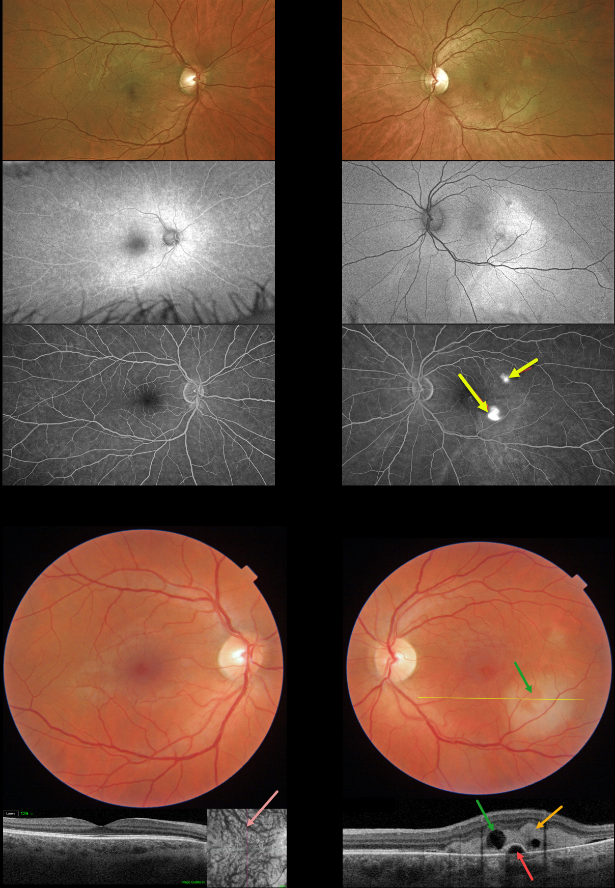
This 42YO male presented with mild symptoms of decreased color saturation and 20/40 vision in his left eye. The right eye was normal with 20/20 vision.
Color imaging shows creamy white subretinal fibrin in the inferotemporal left macula with a smaller area of fibrin superotemporally. Swept-source OCT through the larger inferior lesion shows hyperreflective, subretinal fibrin extending into the outer retinal layers. There is a small underlying retinal pigment epithelial detachment (PED), with a small overlying hyporeflective space corresponding funduscopically to an area devoid of fibrin.
Although the right macula appears normal funduscopically and on B-scan OCT, en face imaging shows somewhat dilated choroidal vessels.
Optos fundus autofluorescence (FAF) shows a hyper-FAF gutter extending from both fibrinous lesions. Fluorescein angiography shows two hyperfluorescent blot leaks.
Learning Points:
In idiopathic central serous chorioretinopathy (ICSC), fluorescein dye diffuses through a physiologic break in the RPE and then pools within the subretinal space. As in this case, the subretinal leak is almost always associated with a PED.
The clearing within the inferotemporal subretinal fibrin, which corresponds to the hyporeflective space within the fibrin on OCT, is caused by serous fluid entering the subretinal space and ‘washing away’ some of the fibrin.
Most patients with ICSC have a single, unilateral leak. Blot leaks are most common, with only 10% of cases exhibiting a smokestack leak. The multifocal leaks and subretinal fibrin noted in our patient are more commonly found in steroid-induced disease (our patient denied steroid exposure). Due to the complete lack of symptoms, initial observation was recommended.



