

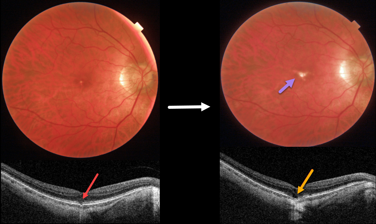
This 58YO male was referred for an asymptomatic foveal lesion in his right eye. There was a history of high myopia prior to LASIK surgery years earlier. Vision was 20/40 OD.
Color photography shows a small atrophic foveal scar. OCT shows a focal EZ defect with a possible tiny choroidal excavation. He returned about 2 years later for a routine follow-up visit.
Although he was binocularly without a new complaint, his vision had decreased to 20/200. There was now a definite, much enlarged foveal lacquer crack. OCT scanning shows a new outer foveal microdefect.
Learning Points:
Outer foveal microdefects (Cohen et al, Ophthalmology Retina 2021;5:553-561) have been described in numerous conditions, including macular telangiectasia, tamoxifen use, ABCA4 disorders (cone-rod dystrophies, Stargardt disease, and fundus flavimaculatus), phototoxicity, trauma, and vitreomacular traction disorders.
We are not aware of a prior report showing these lesions in association with myopic lacquer cracks. These defects, caused by focal loss of the outer retinal layers, leave an optically empty space that can be mistaken for subretinal fluid but is more of an optical gap within the outer retina.
Focal choroidal excavation (FCE), originally described by Jampol et al in 2006, is likely part of the pachychoroid spectrum since it is usually associated with a thickened choroid and pachyvessels. The FCE can be observed, although symptomatic manifestations, including active central serous retinopathy or macular neovascularization, often require treatment. An excellent review is by Verma et al, Br J Ophthalmol 2021;105:1043-1048.



