

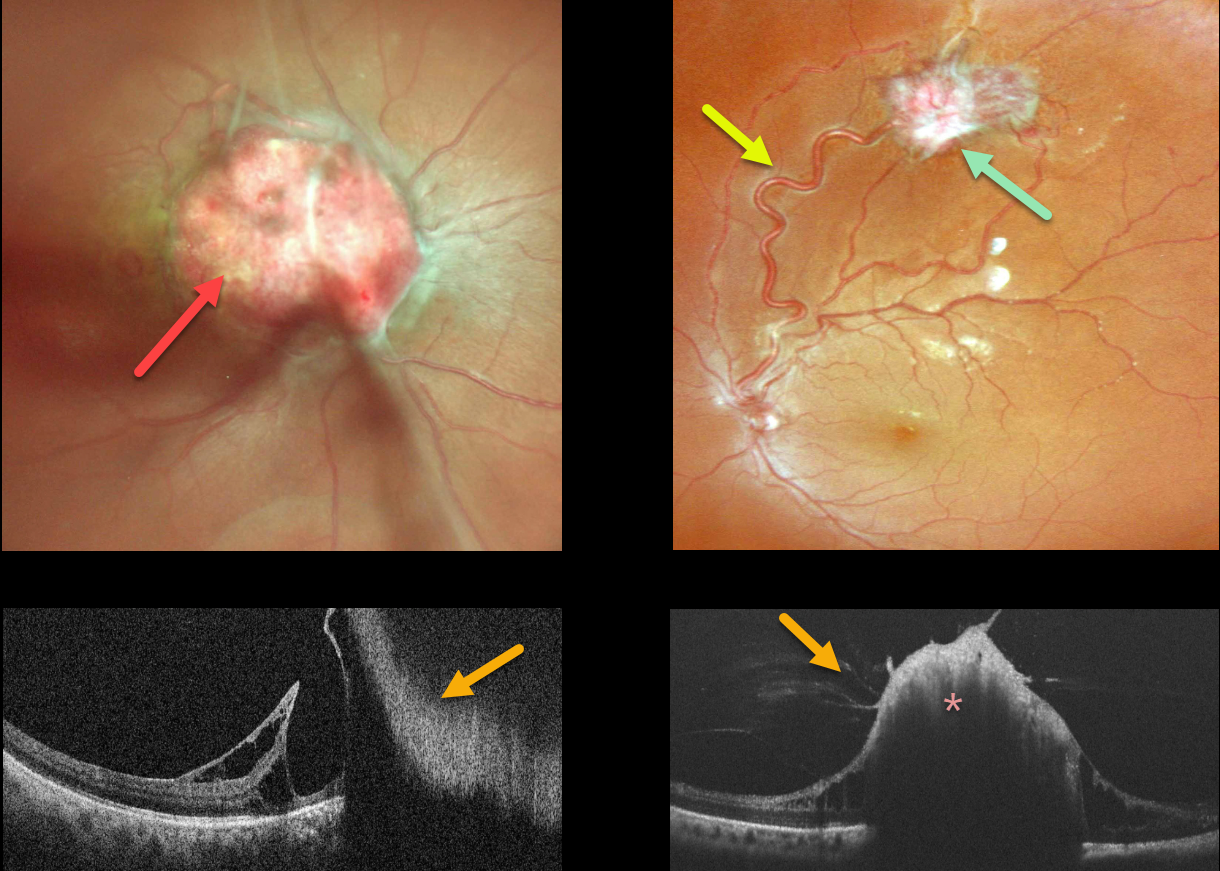
This 31YO healthy female presented with a 3-day history of sudden vision loss in her right eye. Other than a recent history of hypertension, past medical and family history were negative. Her twins both died at 6 months of age from complications related to prematurity. Vision was counting fingers OD and 20/20 OS.
There was a moderate diffuse vitreous hemorrhage in her right eye. Color imaging shows an endophytic capillary hemangioma overlying the right optic nerve. Color imaging of her left eye shows a superotemporal peripheral variably fibrosed retinal capillary hemangioma fed and drained by dilated and irregular vessels.
On OCT, the angiomas appear as thickened, hyperreflective lesions with overlying vitreous traction.
A provisional diagnosis of Von Hippel-Lindau (VHL) disease was made, presumably from a new mutation, and she was referred to neurology and nephrology for further systemic evaluation. Unfortunately, she was subsequently lost to follow-up.
Learning Points:
Retinal capillary hemangiomas are orange-red lesions associated with a prominent pair of feeding and draining vessels and can cause severe vision loss or blindness due to exudative retinal detachment. These benign vascular tumors need to be aggressively treated. Common treatment options include thermal laser, cryotherapy, and anti-VEGF injections.
Retinal capillary hemangioma is a predominant feature of VHL, an autosomal dominant condition caused by mutations in the VHL tumor suppressor gene. Patients develop benign and malignant tumors, including central nervous system hemangioblastoma, pheochromocytoma, and renal cell carcinoma.



