

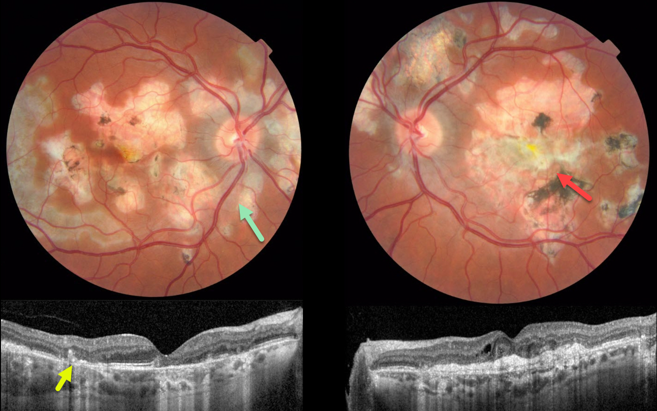
This 34YO male presented with a one-day history of severe vision loss in his left eye. Vision was 20/30 OD and 20/400 OS.
Color imaging shows bilateral areas of centrifugal pseudopod-like projections of atrophic scarring. The peripapillary scarring in the right eye seems to radiate outward from the optic nerve. There is a large, mostly atrophic disciform scar in the left eye, along with bilateral patches of variably pigmented scarring.
OCT scanning shows hyperreflective subfoveal lesions likely representing spontaneously involuted type 2 macular neovascularization (MNV) with overlying disorganized outer retinal layers.
Our patient’s vision in his left eye is remarkably good, given the macular and OCT appearance. It is unclear why he presented with acute vision loss, given the chronic appearance of his findings.
Learning Points:
Relentless placoid chorioretinitis has clinical and angiographic features resembling both serpiginous and APMPPE. Relentless placoid demonstrates the multifocal lesions of APMPPE, with more meandering, less patchy, serpiginous lesions.
Additionally, these patients will have recurrent inflammation typical of serpiginous but not APMPPE. They may also develop MNV that is more typical of the serpiginous form.



