

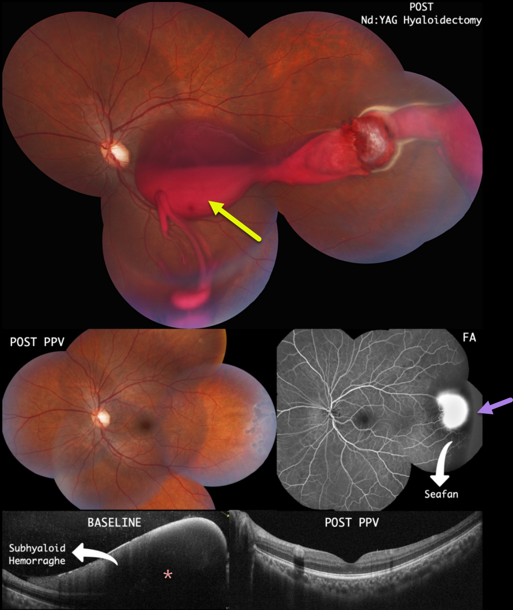
This 22YO male presented with 3 days of vision loss in his left eye. There was a past medical history of Hemoglobin SC Disease. He denied any Valsalva maneuvers. He remembered seeing a retinal doctor several months earlier with the diagnosis of proliferative sickle retinopathy. Vision was counting fingers OS.
Color imaging shows a large subhyaloid/vitreous hemorrhage extending from the temporal midperiphery into the macula. The blood seemed to originate from a probable area of neovascularization (sea fan). OCT scanning confirmed the subhyaloid location of the blood.
Immediately following Nd:YAG laser hyaloidotomy, the blood began to drain into the inferior vitreous. Peripheral scatter laser into the temporal ischemic retina was also performed.
However, he subsequently developed a dense vitreous hemorrhage requiring vitrectomy with endolaser. One month after surgery, the vitreous was clear, the neovascularization had regressed, and the macular appearance had normalized. Vision was 20/20.



